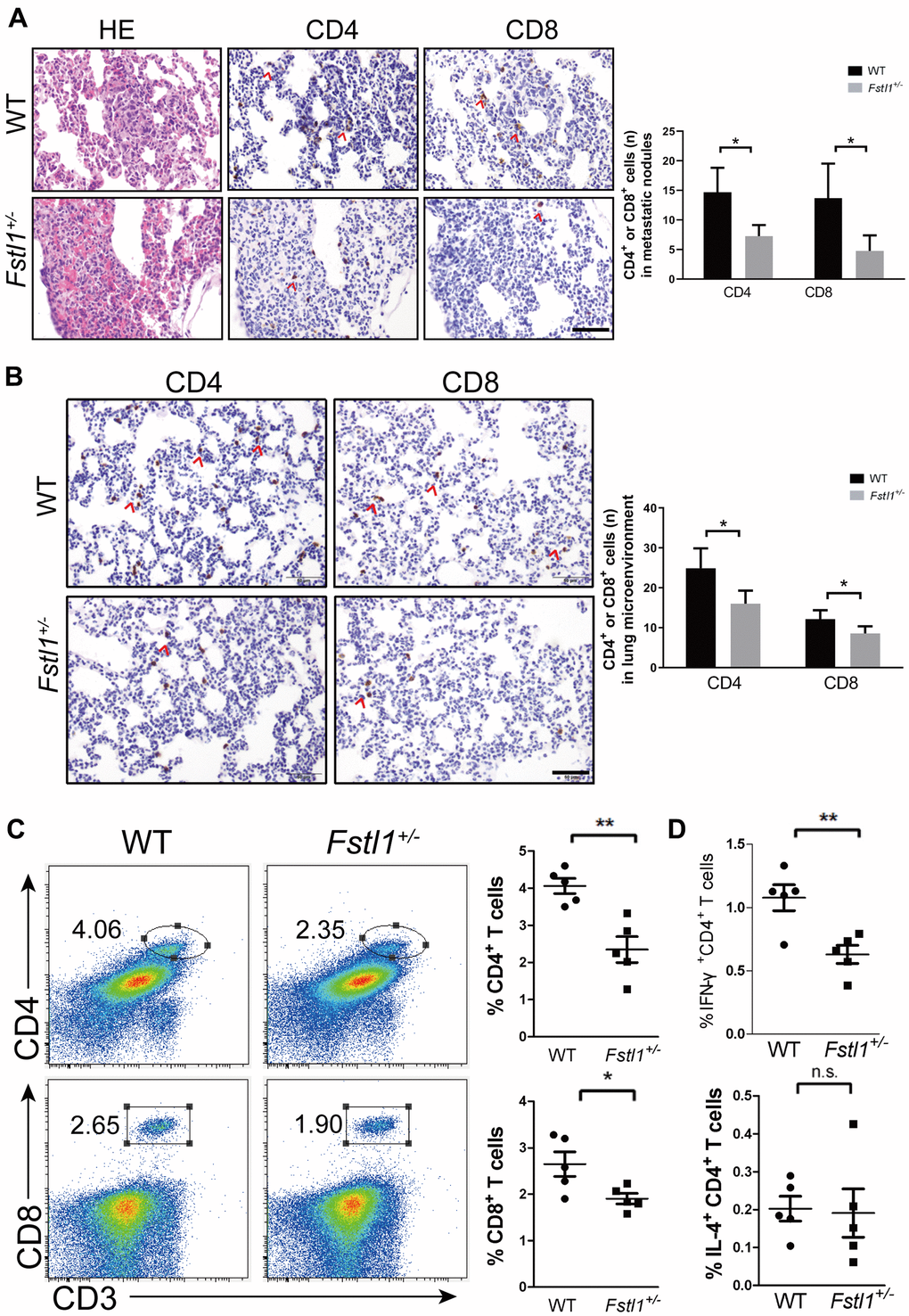
Figure 2.Fstl1+/- mice displayed decreased Th1 and CD8+ T cells in metastatic lungs. (A) H&E staining of metastatic nodules from WT and Fstl1+/- mice. Scale bar, 50 μm. Representative IHC staining of CD4 and CD8 T cells in metastatic nodules from WT and Fstl1+/- mice. Scale bar, 50 μm. The numbers of CD4 and CD8 positive cells in lung metastatic nodules (n=3, WT; n=5, Fstl1+/-). (B) Representative IHC staining of CD4 and CD8 T cells in lung slices of WT and Fstl1+/- tumor-bearing mice. Scale bar, 50 μm. The numbers of CD4 and CD8 positive cells in the lung microenvironment (n=5). (C) Representative flow cytometry profiles presenting the proportions of CD4+ and CD8+ T cells in metastatic lungs of WT and Fstl1+/- mice. Quantification of the proportions of CD4+ and CD8+ T cells within the gated live cells in the metastatic lungs of WT and Fstl1+/- mice (n=5). (D) Quantification of the proportions of IFN-γ+ CD4+ and IL-4+ CD4+ T cells within the gated live cells in the metastatic lungs of WT and Fstl1+/- mice (n=5). Data presented as mean ± SD. Each dot in the graphs represents an individual mouse. n.s., not significant;*p < 0.05, **p < 0.01.