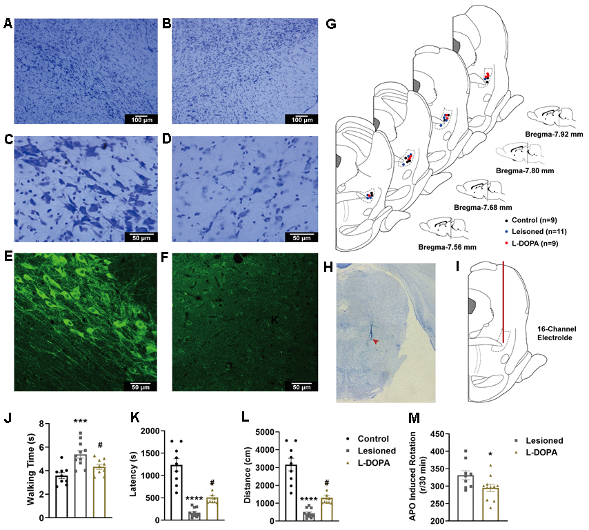
Figure 2.(A–D) Exemplar images of cresyl violet-stained coronal sections of the substantia nigra pars compacta of animals with 6-hydroxydopamine (6-OHDA) lesions. (A) control hemisphere, 100×; (B) lesioned hemisphere, 100×; (C) control hemisphere, 400×; (D) lesioned hemisphere, 400×. (E, F) fluorescence immunohistochemical staining of dopaminergic neurones for tyrosine hydroxylase in the substantia nigra pars compacta. (E) Control hemisphere, 200×; (F) lesioned hemisphere, 200×. (G) Schematic representation of electrode positioning in rats of different groups. (H) Image of a coronal section of PPN stained with cresyl violet. The location of the electrode tip is indicated by a red arrowhead. (I) Schematic representation of the electrode path (red line) into the pedunculopontine nucleus (PPN). (J) Walking time along a control ladder in lesioned and levodopa (L-DOPA) rats. (K) Latency in the rotarod test. (L) distance in the rotarod test. (M) Number of apomorphine (APO)-induced rotations after 30 min in lesioned and L-DOPA rats. * p < 0.05, *** p < 0.001, **** p < 0.0001 compared to the control group; # p < 0.05 compared to the lesioned group.