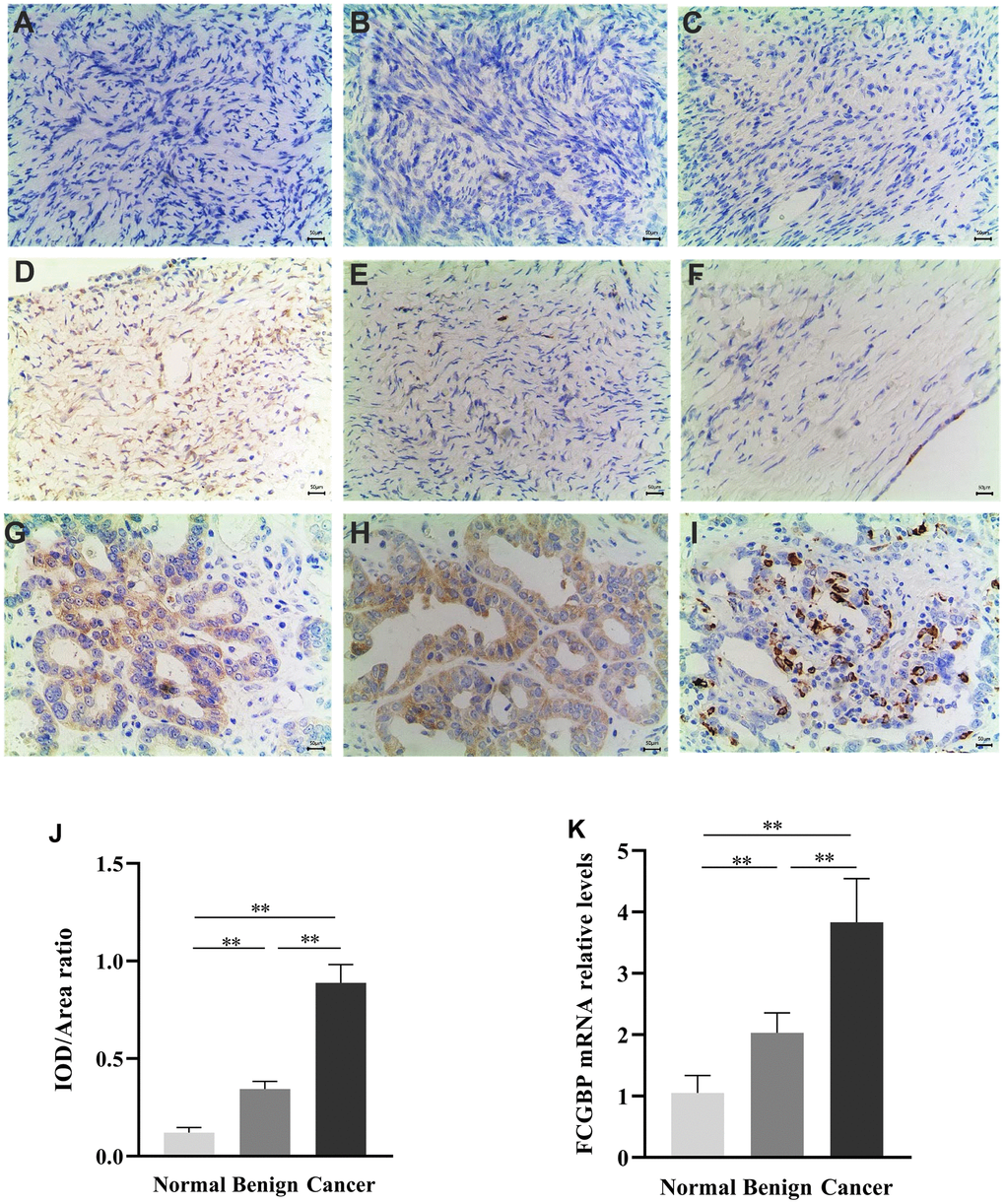
Figure 2.Expression of FCGBP in ovarian cancer tissues. (A–C) Representative images of immunohistochemistry showing FCGBP expression in normal ovarian tissues. (D–F) Representative images of immunohistochemistry showing FCGBP expression in benign ovarian cancer tissues. (G–I) Representative immunohistochemistry images showing FCGBP expression in ovarian cancer tissues. (J) IOD/area ratio of the indicated immunohistochemistry images. (K) qRT-PCR analysis of FCGBP expression in the indicated groups.