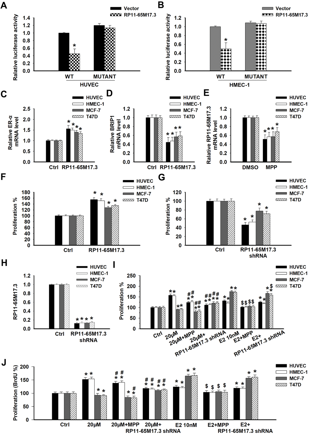
Figure 2.The association among RP11-65M17.3, BRIP1 and ERα in ECs and BCCs and the involvement of RP11-65M17.3 and ERα in calycosin mediated regulation of cell proliferation. (A, B) A luciferase reporter assay was performed to determine whether BRIP1 is the downstream target of RP11-65M17.3 in HUVECs and HMEC-1 cells. *p < 0.05 vs. vector. (C–E) The existence of a positive feedback loop between RP11-65M17.3 and ERα was demonstrated in HUVECs, HMEC-1 cells, MCF-7 cells and T47D cells. The cells were treated with the plasmid construct pCDNA3.1-RP11-65M17.3 or the ERα antagonist MPP. The mRNA expression levels of ERα, BRIP1, and RP11-65M17.3 were determined by qRT-PCR, and β-actin was used as the internal control. (F, G) The overexpression of RP11-65M17.3 stimulated cell proliferation, while the knockdown of RP11-65M17.3 inhibited proliferation, as detected by CCK8 assay. *p < 0.05 vs. control. (H) Pretreatment with pCDNA3.1-RP11-65M17.3 shRNA downregulated the expression of RP11-65M17.3 in HUVECs, HMEC-1 cells, MCF-7 cells and T47D cells. *p < 0.05 vs. control. (I, J) The four cell lines were then treated for 48 h with 20 μM calycosin, 20 μM calycosin plus MPP, 20 μM calycosin plus RP11-65M17.3 shRNA, 10 nM E2, 10 nM E2 plus MPP, or 10 nM E2 plus RP11-65M17.3 shRNA. Cell proliferation was analyzed using the CCK-8 assay and BrdU assay. Representative data from three independent experiments are shown. *p < 0.05 vs. control (0 μM); #p < 0.05 vs. 20 μM calycosin alone; $p < 0.05 vs. 10 nM E2 alone.