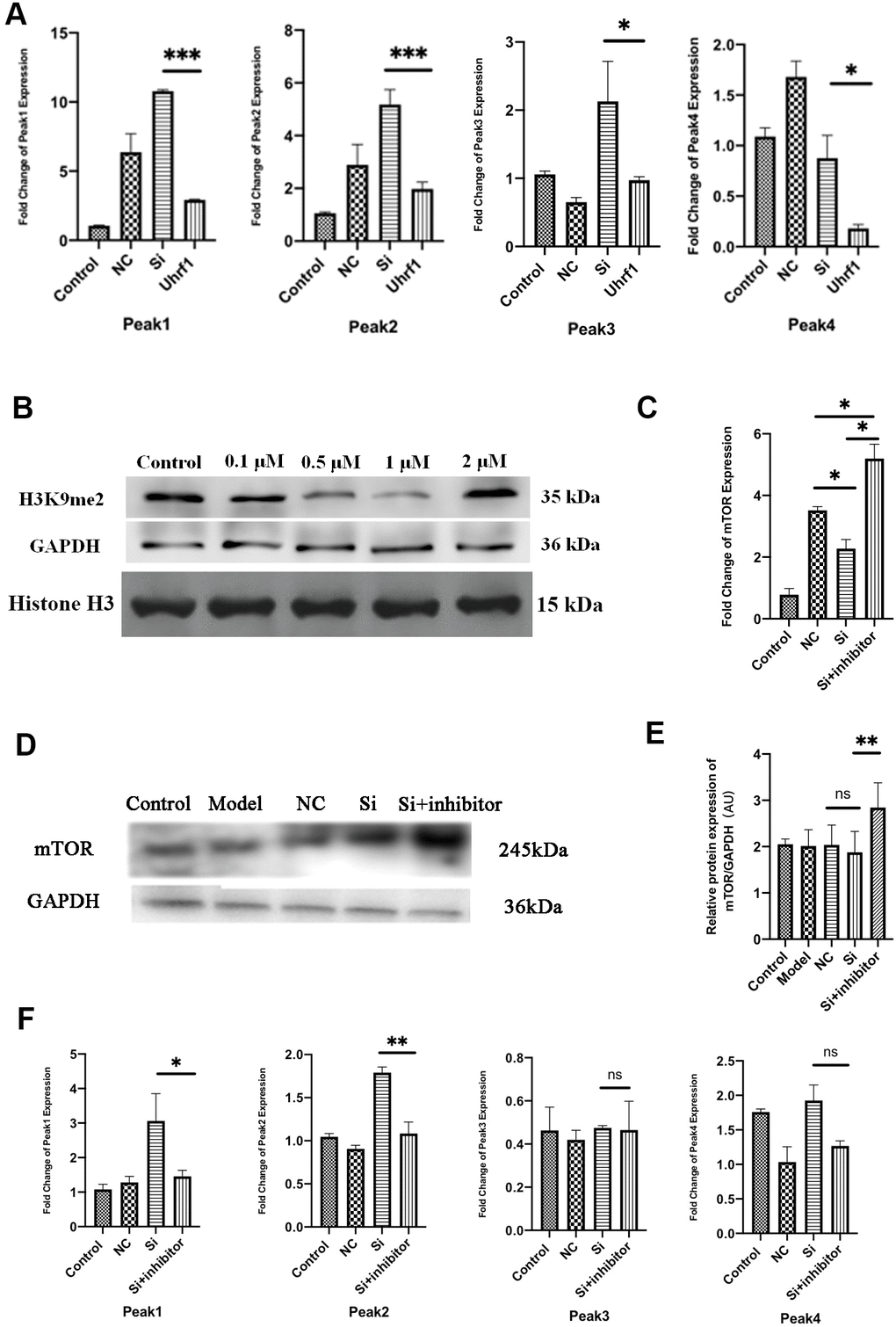
Figure 6.Uhrf1 regulates mTOR expression through H3K9me2. (A) The relative mRNA expressions of Peak1-4 in Myocardial ischemia-reperfusion model in vitro were determined by qRT-PCR. (B) Western blot was used to detect the inhibitory effect of G9a inhibitor on H3K9me2 in cardiomyocytes by different concentrations (0.1 µM, 0.5 μM, 1.0 μM and 2.0 μM). GAPDH and the total histone H3 were used as loading controls. (C) The relative mRNA expressions of mTOR in myocardial ischemia-reperfusion model in vitro were determined by qRT-PCR after adding 1.0 μM G9a inhibitor to Si group. (D) Western blot was used to detect the expression level of mTOR protein after adding 1.0 μM G9a inhibitor to Si group. GAPDH serves as a loading control. (E) Expression of mTOR protein relative to GAPDH data from 3 biological repeats is shown. (F) The relative mRNA expressions of Peak1-4 in Myocardial ischemia-reperfusion model in vitro were determined by qRT-PCR after adding 1.0 μM G9a inhibitor to Si group. Data shown are mean ± SD. *P < 0.05, **P < 0.01, ***P < 0.001, ****P < 0.0001. N=3 per group. Model, in vitro oxidative stress model; NC, negative control of RNAi; si, RNAi knockdown of Uhrf1; Uhrf1, Uhrf1 overexpression.