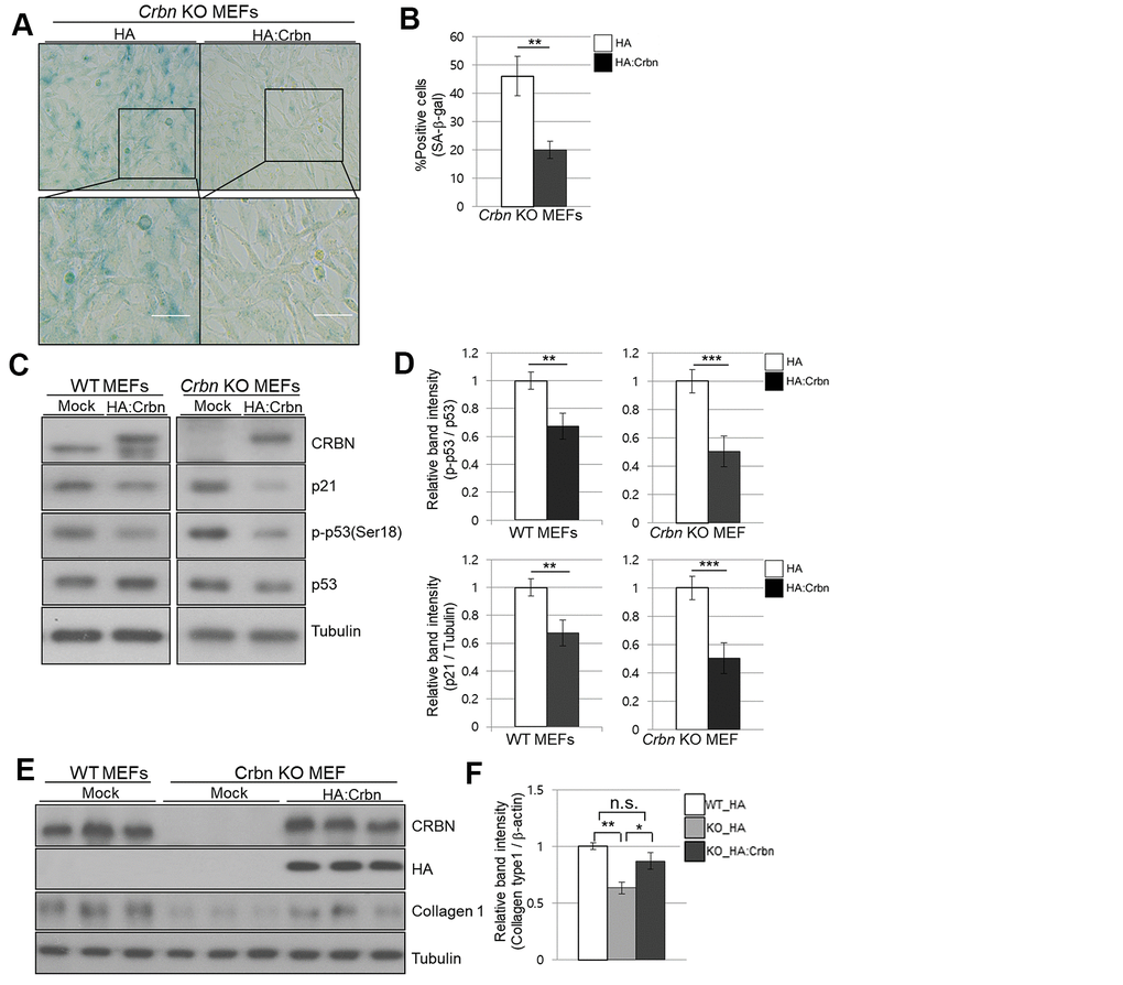
Figure 6.Ectopic overexpression of CRBN clears the SA-β-Gal signal and recovers the protein level of Collagen I in cultured fibroblast. (A) Representative staining images showing SA-β-Gal (blue-stained cells) along with the overexpression of CRBN in primary MEFs. Scale bar = 100μm. (B) Quantification of SA-β-Gal-positive cells shown in (A). Results are expressed as the percentage of stained cells (mean+SEM). The results shown are representative of four independent experiments. (C) The WT and CRBN KO primary MEFs were transiently transfected with HA: CRBN or empty vector. Cells were harvested after 24h and the protein lysates were subjected to immunoblotting with the anti-CRBN, anti-p53, anti-p-p53(Ser18), anti-p21, and anti–Tubulin antibodies. (D) The relative band ratio as determined by densitometric analysis of the blots in (C). (E) Cell lysates were prepared from WT and CRBN KO primary MEFs transfected with HA: CRBN or empty vector. Western blots of the protein lysate were probed with the anti-CRBN, anti-HA, anti-collagen I, and anti–Tubulin antibodies. Tubulin was used for equal protein loading. (F) The relative band intensity was measured by densitometric-analysis of the blots in (E). The results shown are representative of five independent experiments. *P < 0.05; **P < 0.01; ***P < 0.005; n.s., not significant.