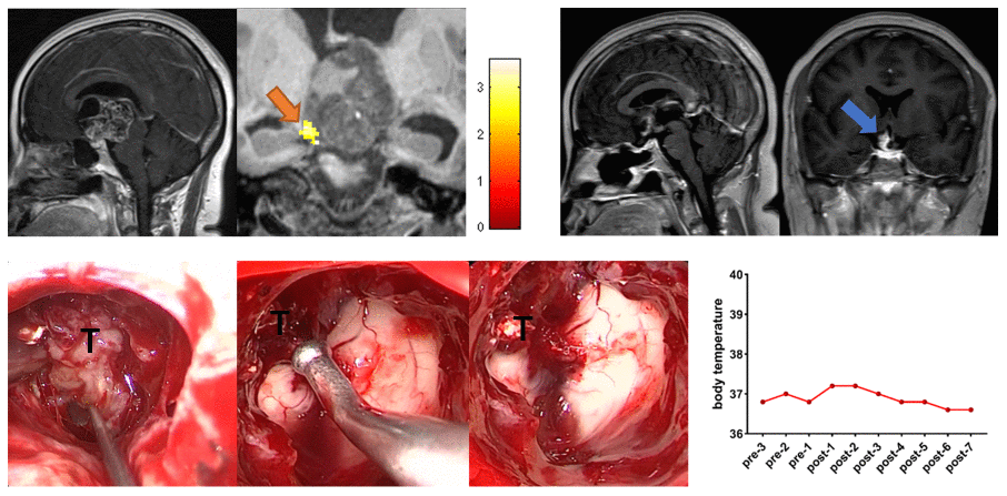
Figure 3.Preoperative midsagittal contrast-enhanced T1-weighted MRI scans revealed a large solid tumor extending up into the third ventricle, and the coronal task-related fMRI showed the activated POAH of the hypothalamus in case 3. The orange arrow indicated the activated POAH. The postoperative sagittal and coronal contrast-enhanced T1-weighted MRI showed that NTR was achieved. The blue arrow indicated the tumor that unremoved. Intraoperative photographs showed that the tumor adhered tightly to the surrounding tissues, especially the POAH of the hypothalamus, which was localized preoperatively. To protect the function of the hypothalamus, the part of the tumor that adhered closely to the hypothalamus was not forcibly removed. No significant increase in body temperature was observed during the perioperative period. fMRI: functional magnetic resonance imaging; POAH: preoptic and anterior hypothalamic region; NTR: near-total resection; T: tumor.