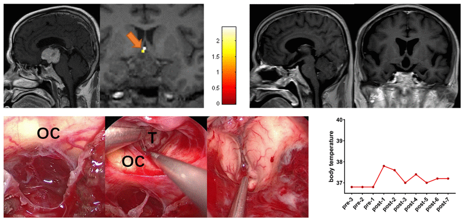
Figure 4.Preoperative midsagittal contrast-enhanced T1-weighted MRI scans showed a large solid tumor involving the third ventricle, and the coronal task-related fMRI showed the activated POAH of the hypothalamus in case 12. The orange arrow indicated the activated POAH. The postoperative sagittal and coronal contrast-enhanced T1-weighted MRI showed that GTR was achieved. Intraoperative photographs showed that the tumor was removed through the CPC approach combined with the EETLT approach. After complete resection of the tumor, the midbrain aqueduct and hypothalamus could be observed. No significant increase in body temperature was observed during the perioperative period. fMRI: functional magnetic resonance imaging; POAH: preoptic and anterior hypothalamic region; GTR: gross total resection; CPC: chiasm-pituitary corridor; EETLT: endoscopic endonasal trans-lamina terminalis approach; OC: optic chiasma; T: tumor.