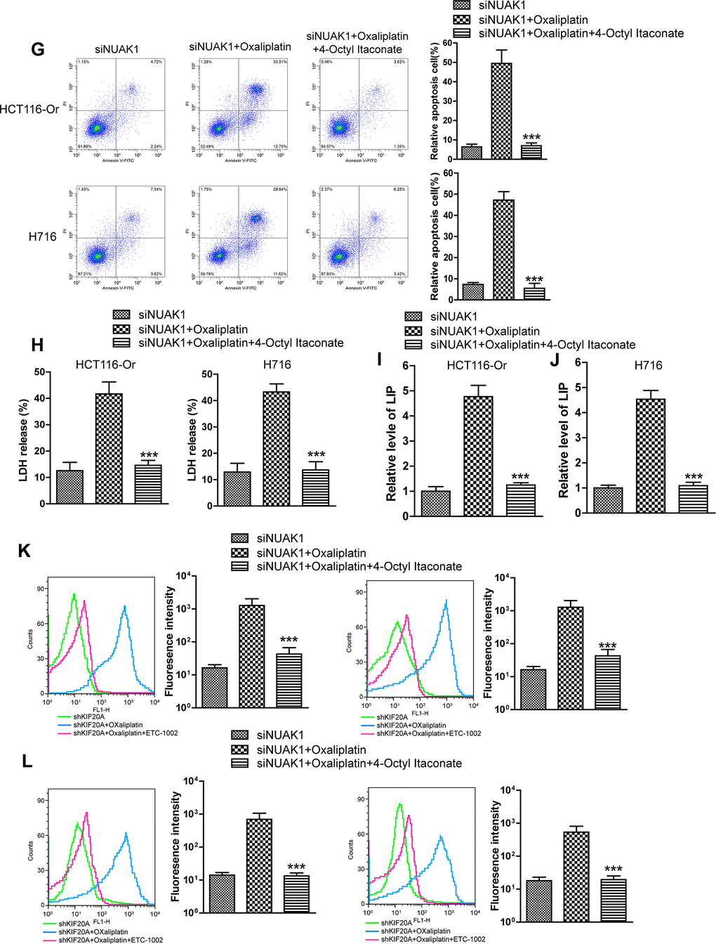
Figure 4.KIF20A/NUAK1 induce the resistance of CRC resistant cell lines to Oxaliplatin through activating Nrf2 pathway. (G) Cell death was assessed by flow cytometry (annexin V-FITC/PI staining) to observe whether 4-Octyl Itaconate would affect the lethal effect of oxaliplatin on NUAK1-silenced colorectal cancer cells in vitro. Left, representative results of annexin V-FITC/PI staining. Right, quantitative analysis. Top, HCT116-Or cells. Bottom, H716 cells. The data are presented as the mean ± SD, ***p < 0.001 (versus siNUAK1+Oxaliplatin). (H) Cell death was assessed by LDH release assay to observe whether 4-Octyl Itaconate would affect the lethal effect of oxaliplatin on NUAK1-silenced colorectal cancer cells in vitro. Top, HCT116-Or cells. Bottom, H716 cells. The data are presented as the mean ± SD, ***p < 0.001 (versus siNUAK1+Oxaliplatin). (I, J) The cellular LIP was analyzed with a flow cytomete to observe whether 4-Octyl Itaconate would affect the LIP induction of oxaliplatin on NUAK1-silenced colorectal cancer cells. (I) HCT116-Or cells. (J) H716 cells. The data are presented as the mean ± SD, ***p < 0.001 (versus siNUAK1+Oxaliplatin). (K, L) The cellular level of ROS (K) and lipid peroxidation (L) was assessed by flow cytometry to observe whether 4-Octyl Itaconate would affect the oxidative damage induction of oxaliplatin on NUAK1-silenced colorectal cancer cells. Left, HCT116-Or cells. Right, H716 cells. The data are presented as the mean ± SD, ***p < 0.001 (versus siNUAK1+Oxaliplatin).