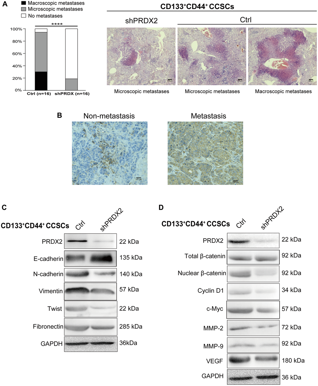
Figure 5.PRDX2 knockdown inhibits the metastatic capacity of CCSCs. (A) Histological analysis of the liver for metastatic lesions was performed by hematoxylin and eosin staining. The metastatic status of the mice (n = 16 per group) with orthotopic implantation of 1 × 10⁴ cells dissociated from HCT116/HT29-control or -shPRDX2 CD133+CD44+ CCSCs is provided separately as macroscopic (black) and microscopic (gray) evidence for metastasis (left panel). Statistical analysis: Fisher’s exact test, ***p < 0.0001. Representative images are from two mice receiving control-HCT116-CD133+CD44+ CCSCs and a mouse receiving shPRDX2-HCT116-CD133-CD44- CCSCs (right panel). (B) Representative IHC staining for PRDX2 reveals that higher PRDX2 expression was detected in the orthotopic tumor tissues from metastatic cases than in tumor samples from nonmetastatic mice. (C) Western blot analysis of EMT protein expression in lysates of control- and shPRDX2-CD133+CD44+ CCSCs. (D) Western blot analysis of Wnt/β-catenin signaling pathway protein expression in lysates of control- and shPRDX2-CD133+CD44+ CCSCs.