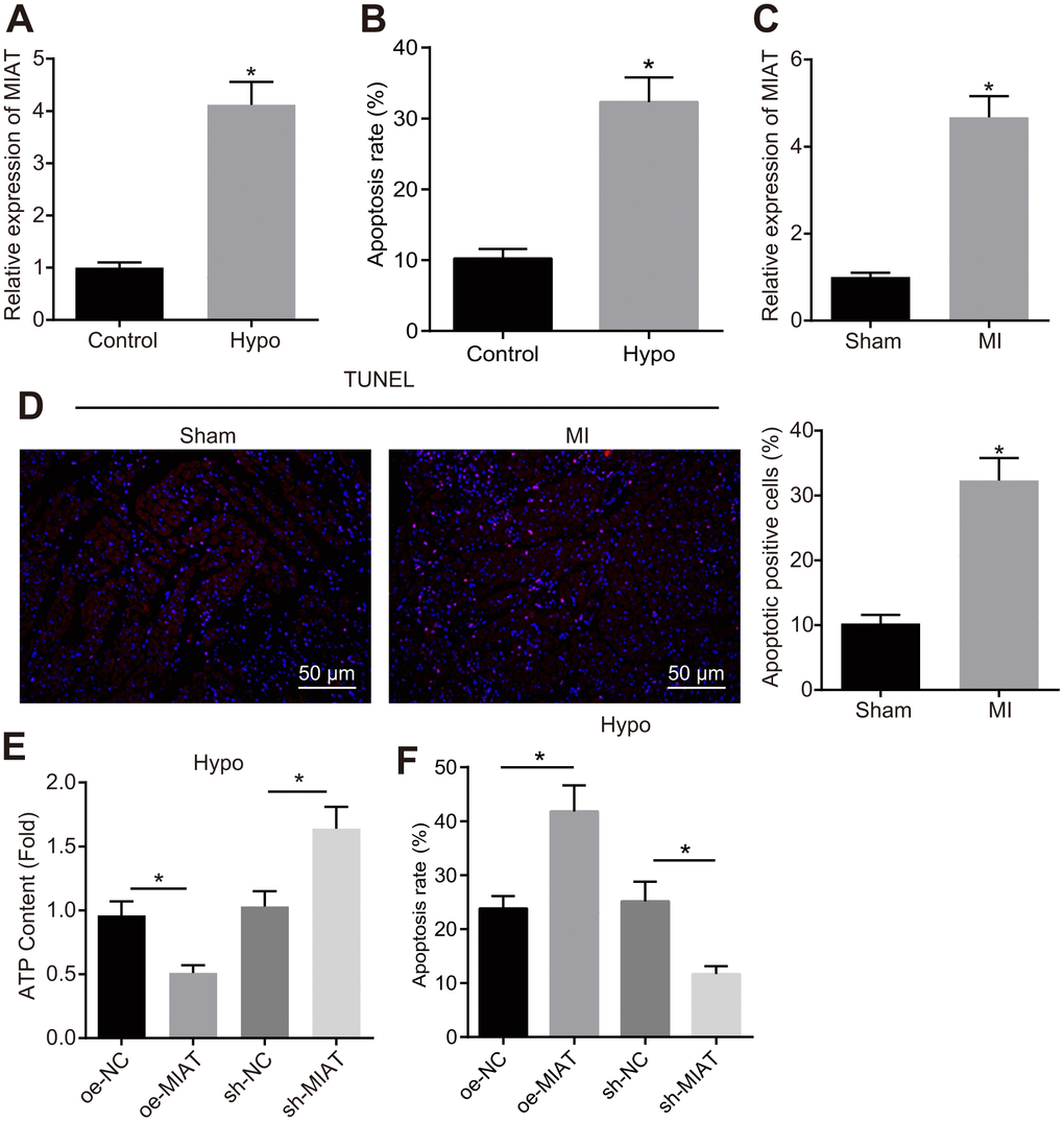
Figure 1.Upregulation of MIAT in hypoxic cardiomyocytes and myocardial tissues from the MI mice. (A) MIAT expression pattern in the cardiomyocytes determined by RT-qPCR normalized to GAPDH. (B) The cardiomyocyte apoptosis detected by flow cytometry. (C) RT-qPCR determination of MIAT expression pattern in the cardiomyocytes in mice 28 d after MI modeling (n = 10). (D) The apoptosis of cardiomyocytes in mice 28 d after MI modeling showed by TUNEL staining (scale bar = 50 μm). (E) The ATP content in hypoxic cardiomyocytes after alteration of MIAT. (F) The apoptosis of hypoxic cardiomyocytes after alteration of MIAT analyzed by flow cytometry. * p < 0.05. The above data were all measurement data, and expressed as mean ± standard deviation. The unpaired t test was used for comparison between two groups, and one-way ANOVA was applied for comparison between multiple groups followed by Tukey’s post hoc test. All the data was collected from 3 independent experiments respectively.