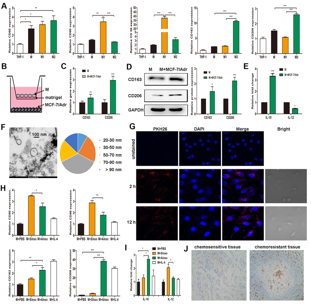
Figure 1.A/exo derived from MCF-7/Adr cells induce macrophages M2 polarization. (A) Expressions of CD68 (macrophage marker), CD80 and CD86 (M1 phenotype markers), CD163 and CD206 (M2 phenotype markers) were analyzed in THP-1 monocytes and the derived macrophages. (B) Macrophages (M) were incubated with MCF-7/Adr cells in a co-culture chamber to avoid direct cell contact. (C) After 48 h, M2 phenotype markers CD163 and CD206 were analyzed by PCR. (D) CD163 and CD206 were analyzed by western blot. (E) IL-10 and IL-12 expression levels in macrophages incubated with MCF-7/Adr cells were evaluated by ELISA. (F) Representative transmission electron microscopic image of A/exo showing a spheroid shape with size ranging from 30 to 90 nm (bar indicates 100 nm). (G) Representative fluorescence microscopic images showing the uptake of unstained A/exo or PKH26-labeled A/exo (red) by macrophages (blue). (H) Macrophages were incubated with A/exo, S/exo, or controls (PBS and IL-4) for 48 h, and expressions of CD80, CD86, CD163, and CD206 were analyzed by PCR. Macrophages treated with IL-4 was used as a positive control. (I) IL-10 and IL-12 expression levels in macrophages incubated with A/exo, S/exo, or controls (PBS and IL-4) were evaluated by ELISA. (J) Expression of CD163 (grey) was examined by IHC in chemoresistant breast cancer tissues and chemosensitive samples (magnification × 200). Data are shown as mean ± SD, n = 3 independent experiments; * P<0.05, ** P<0.01, and *** P<0.001 compared with controls.