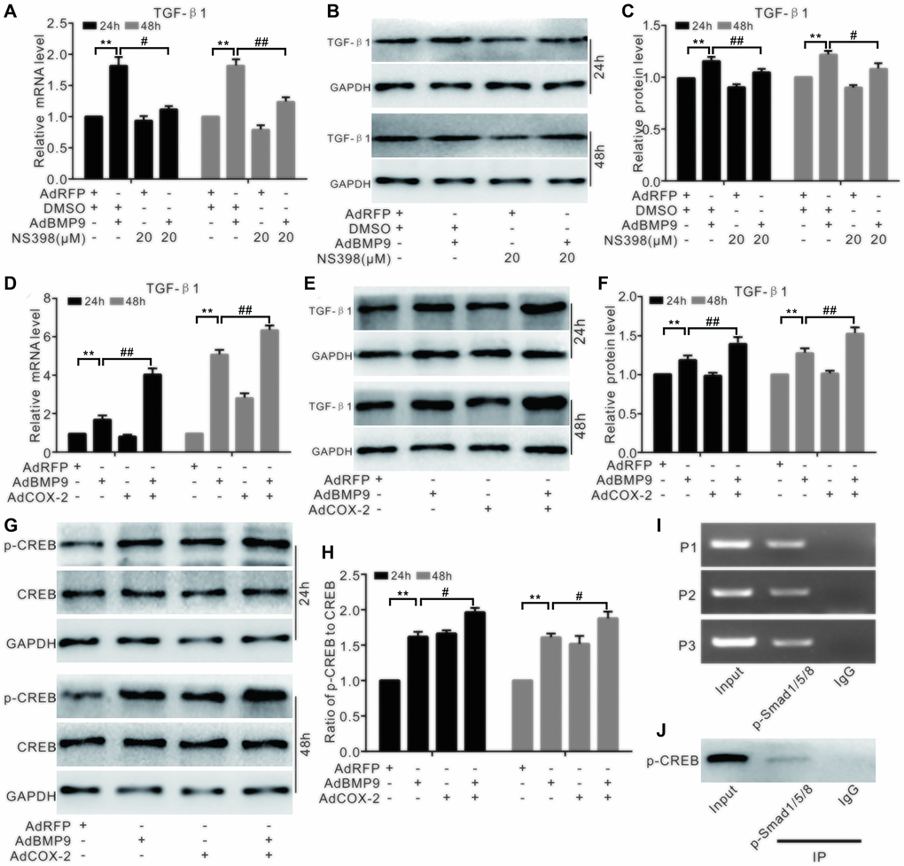
Figure 7.The effect of COX-2 and BMP9 on TGF-β1 expression in C3H10T1/2 cells. (A) Real-time PCR analysis shows the mRNA level of TGF-β1 was affected by BMP9 and/or NS-398. (B) Western blotting shows that the level of TGF-β1 was affected by BMP9 and/or NS-398. (C) Quantification of western blots shows that TGF-β1 level was affected by BMP9 and/or NS-398. (D) Real-time PCR assay shows that TGF-β1 mRNA expression was affected by BMP9 and/or COX-2. (E) Western blotting shows that TGF-β1 level was affected by BMP9 and/or COX-2. (F) Quantification of the western blots shows that TGF-β1 level was affected by BMP9 and/or COX-2. (G) Western blotting shows that the level of CREB and p-CREB was affected by BMP9 and/or COX-2. (H) Quantification of the western blots shows that the levels of CREB and p-CREB was affected by BMP9 and/or COX-2. (I) ChIP assay shows the enrichment of p-Smad1/5/8 at the TGF-β1’s putative promoter region. (J) IP assay shows p-CREB may interact with p-Smad1/5/8. NS-398: COX-2 specific inhibitor; “**”p < 0.01, “#”p < 0.05, and “##”p < 0.01.