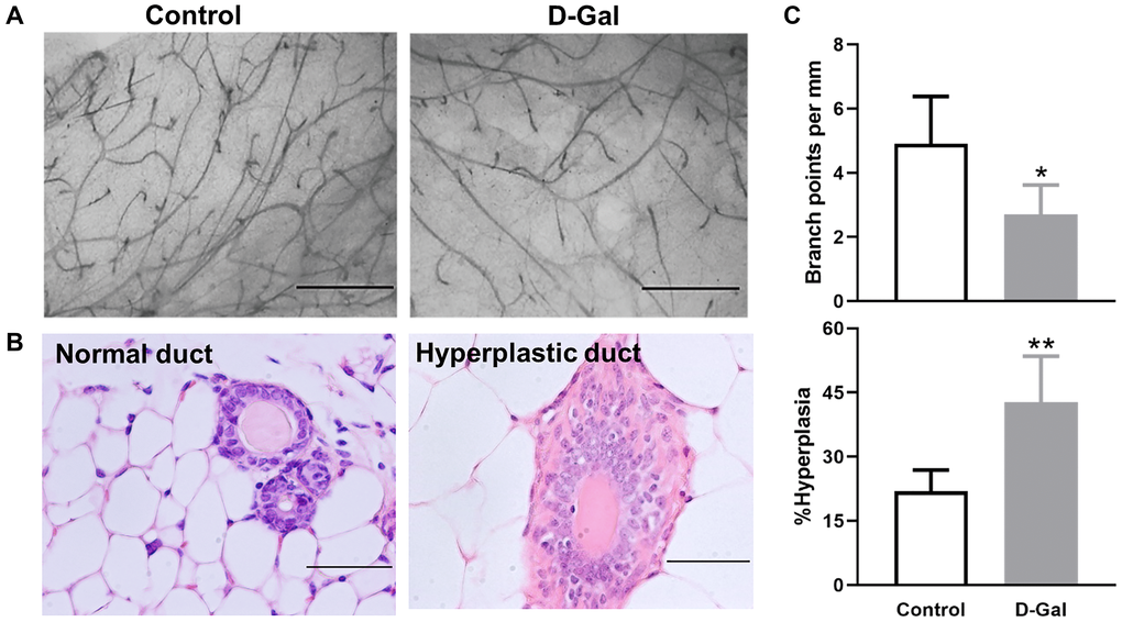
Figure 2.D-galactose affects mammary gland morphology and pathology. (A) Representative examples of whole mount carmine alum staining of mammary glands from control and D-galactose-treated mice (scale bars, 1 mm); (B) H&E histological images showing normal and hyperplastic ducts from control and D-galactose-treated mice (scale bars, 100 μm); (C) Quantification of branch points per millimeter (mm) duct (upper panel) and percent of hyperplastic ducts (lower panel) in control and D-galactose-treated mice (n = 5). Asterisks, significant difference between control and D-galactose (*P < .05, **P < .01).