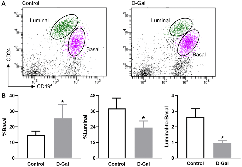
Figure 3.D-galactose alters mammary basal/luminal cell pools. (A) Representative flow cytometry analysis of mammary epithelial cells from control and D-galactose-treated mice. Basal cells express high levels of CD49f, and luminal cells express high levels of CD24; (B) Quantification of % basal cell, % luminal cell, and luminal-to-basal cell ratio in mammary epithelial cells isolated from control and D-galactose-treated mice (n = 5). Asterisks, significant difference between control and D-galactose (*P < .05).