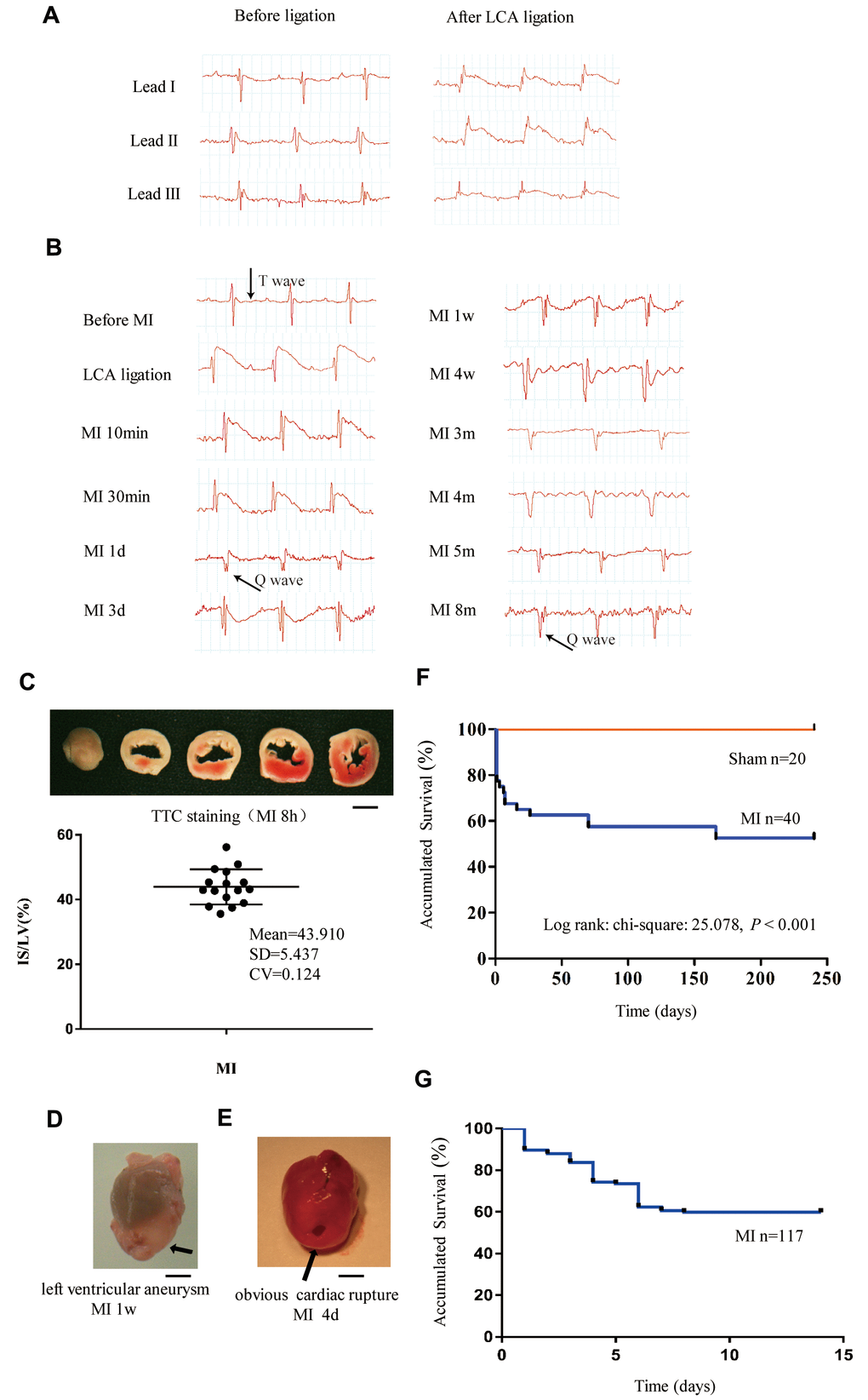
Figure 1.Monitoring of long-term MI mouse model for 8 months. (A) Limb leads I, II and III of electrocardiogram (ECG) reliably reflect the acute ischemia induced by left coronary ligation in C57BL/6 mice. (B) Lead II ECG can reflect different stages of myocardial infarction (MI) for a time period of 8 months. (C) Triphenyl tetrazolium chloride (TTC)-stained sections of heart in mice with MI for 8 h; the infarct size (IS)/ left ventricle (LV) was approximately 44% in the MI groups. (D) Representative left ventricular aneurysm formed 1 week after MI; (E) Example necropsy images of a dead mouse showing obvious rupture in the left ventricle free wall. (F): Kaplan–Meier survival analysis of mice subjected to MI or sham operation for 8 months. (G) Kaplan–Meier survival analysis of mice subjected to MI or sham operation for 14 days, n = 114 in MI group; Scale bar =2 mm for panel (C, D and E).