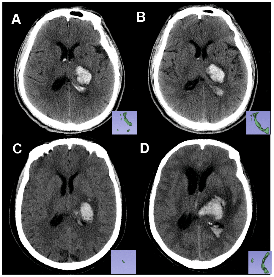
Figure 4.Non-contrast CT images of two patients with similar hematoma but different experiences. Pictures in the lower right corner are the 3-D images of the IVH. Image (A, B) are the baseline and follow-up CT images, respectively, of patient A: a 61-year-old male who had a Rad-score of -2.2119681 (<-1.7259179). Image (C, D) are the baseline and follow-up CT images, respectively, of patient B: a 67-year-old female with a Rad-score of -1.6176548 (>-1.7259179). Within 24 hours from symptom onset, the IVH volume of patient A changed from 5.62 mL to 6.84 mL, and the IVH volume of patient B changed from 0.5 mL to 12.4 mL. Patient B experienced IVH growth while patient A did not.