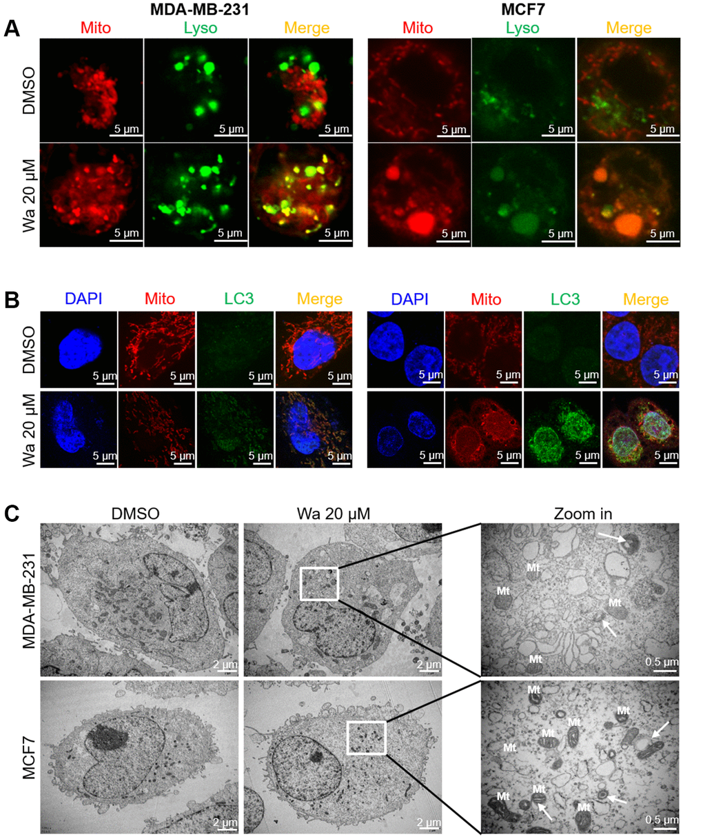
Figure 5.Warangalone induces cell mitophagy. MDA-MB-231 and MCF7 cells were treated with 20 μM warangalone for 12 h. A CLSM was used to observe co-staining. (A) Co-staining of lysosomes (LysoTracker™ Green DND) and mitochondria (MitoTracker™ Red CMXRos). (B) Co-staining of LC3 (labeled with Alexa Fluor 488) and mitochondria (MitoTracker™ Red CMXRos). (C) Transmission electron microscopy was used to observe mitophagy. Nuclei labeled with DAPI. Arrows indicate mitophagy.