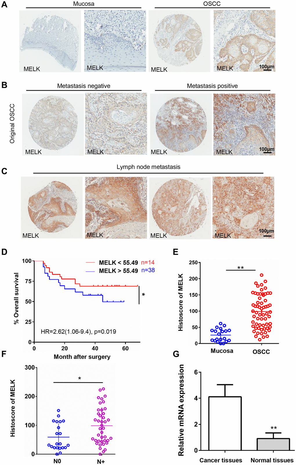
Figure 1.High MELK expression in OSCC tissues correlates closely with lymph node condition. (A) Representative images of MELK expression assessed via IHC staining normal oral mucosa (left) and OSCC (right). Scale bars = 100 μm. (B) Typical images of MELK expression assessed via IHC staining in metastasis-negative (left) and metastasis-negative OSCC (right). Scale bars = 100 μm. (C) Representative photographs of MELK in lymph node metastasis. Scale bars = 100 μm. (D) Kaplan–Meier analysis of OSCC patients with MELK expression level. (E) Score of MELK in oral mucosa (n = 20) and OSCC (n = 62). (F) Score of MELK in OSCC with or without lymph node metastasis. (G) The relative MELK mRNA level in patients with OSCC paired cancer and normal tissues. *P < 0.05; **P < 0.01; ***P < 0.001.