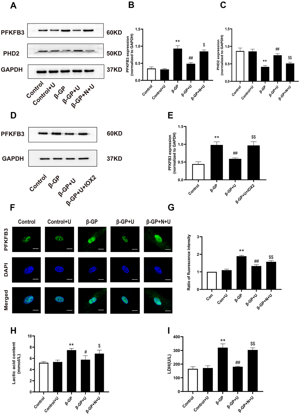
Figure 3.Effects of κ-OR stimulation on the expression of PFKFB3, PHD2, and glycolysis products in VSMCs treated with β-GP. (A) Representative blot images of PFKFB3 and PHD2. (B) Quantitative analysis of PFKFB3 protein expression using densitometry. (C) Quantitative analysis of PHD2 protein expression using densitometry. (D) Representative blot images of PFKFB3. (E) Quantitative analysis of PFKFB3 protein expression using densitometry. (F) After various treatments, PFKFB3 nuclear translocation was evaluated via immunofluorescence using confocal microscopy. At least 10-15 cells per condition were imaged. Scale bar = 10μm. (G) Quantification of PFKFB3 immunofluorescence intensity. (H, I) Lactic acid content and LDH levels were detected. U, U50,488H; β-GP, β-Glycerophosphate disodium salt pentahydrate; N, nor-BNI; Data obtained from quantitative densitometry were presented as means ± SEM. n=5 in each group. **P < 0.01 versus the control group, #P < 0.05 versus the β-GP group, ##P < 0.01 versus the β-GP group, $P < 0.05 versus the β-GP+U group, $$P < 0.01 versus the β-GP+U group.