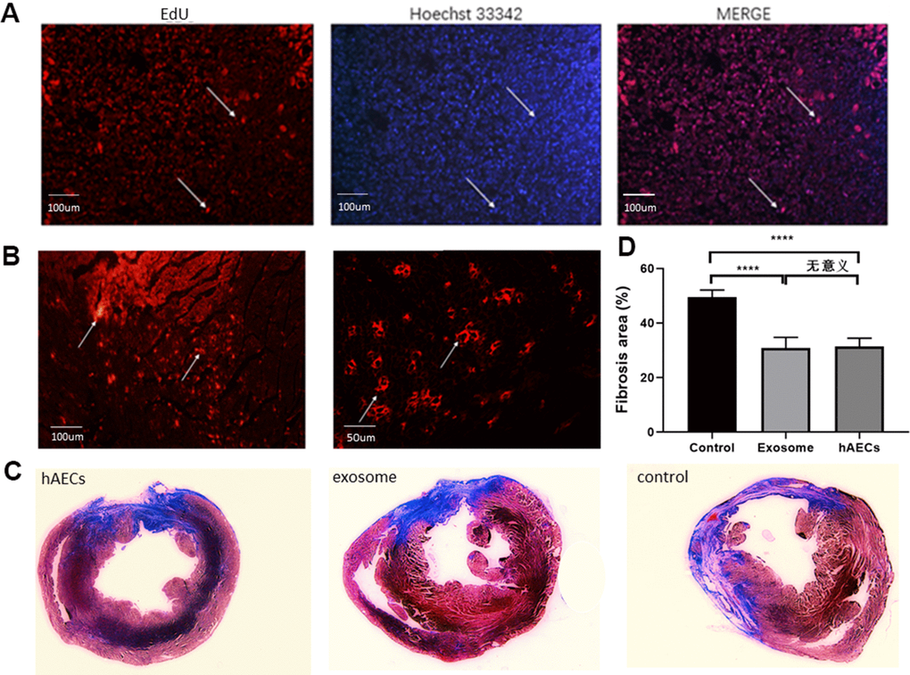
Figure 5.hAECs and exosomes implanted in the heart decrease the fibrosis area. (A) Immunofluorescence of hAECs in vivo 7 days after MI are shown. (B) Immunofluorescence of exosomes in vivo 7 days after MI are shown. (C) Representative images of four consecutive myocardial slices stained with Masson’s trichrome in the hAEC, exosome and control groups 28 days after MI are shown. (D) The bar graph shows quantitative analysis of the LV fibrosis area. The data are shown as the means ± SD, n=4, *P<0.05.