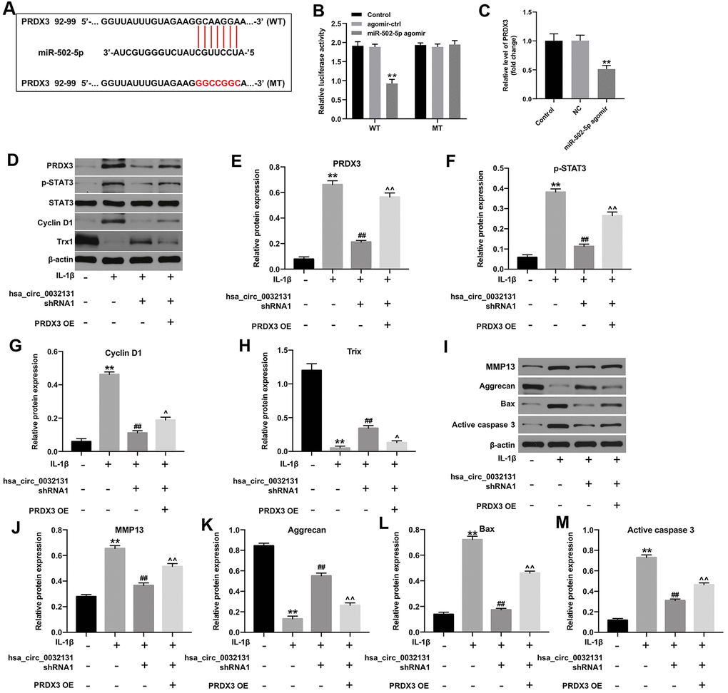
Figure 5.Hsa_circ_0032131 shRNA1 suppressed OA progression by inhibiting the miR-502-5p/PRDX3/Trx1/STAT3 axis. (A) Gene structure of PRDX3 indicated the predicted target site of miR-502-5p in its 3'UTR. (B) The luciferase activity was measured in CHON-001 cells using the dual-luciferase reporter assay following co-transfection with WT/MT PRDX3 3′-UTR plasmid and miR-502-5p. (C) The expression of PRDX3 mRNA in CHON-001 cells was detected by RT-qPCR. (D) The expression of PRDX3, p-STAT3, STAT3, Cyclin D1, and Trx1 in CHON-001 cells was detected by western blotting. (E) The relative expression of PRDX3 was quantified by normalizing it to that of β-actin. (F) The relative expression of p-STAT3 was quantified by normalizing it to that of β-actin. (G) The relative expression of cyclin D1 was quantified by normalizing it to that of β-actin. (H) The relative expression of Trx1 was quantified by normalizing it to that of β-actin. (I) The expressions of MMP13, Aggrecan, Bax and active caspase 3 in CHON-001 cells were detected by western blotting. (J–M) The relative expressions of MMP13, Aggrecan, Bax and active caspase 3 in cells were quantified via normalization to β-actin. **p < 0.01 compared with the control. ##p < 0.01 compared with IL-1β. ^p < 0.05, ^^p < 0.01 compared with IL-1β + hsa_circ_0032131 shRNA1.