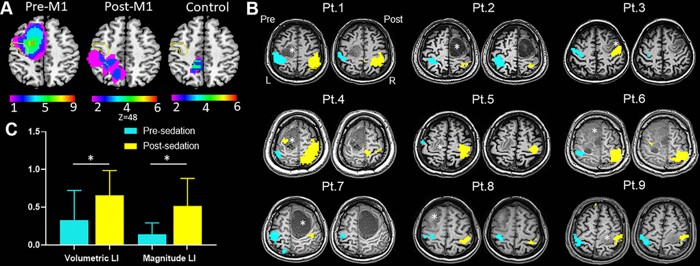
Figure 2.Results of task-fMRI in M1 group. (A) Probability maps of lesion distribution for M1, post-M1 and control groups. The yellow circle denoted location of anterior central gyrus. (B) Hand motor task-fMRI results of nine patients from M1 group. Axial individual anatomical images with superimposed functional activation pre- and post-administration of dexmedetomidine were presented. In the lesional hemisphere, activation of the hand task decreased significantly after sedation. Right (R) and left (L) hemispheres are marked. *indicates locations of the lesions. (C) The bar graph showed both magnitude lateralization index (LI-M) and volumetric LI (LI-V) of M1 increased significantly after sedation in M1 group (mean with 95% CI) (*P < .05).