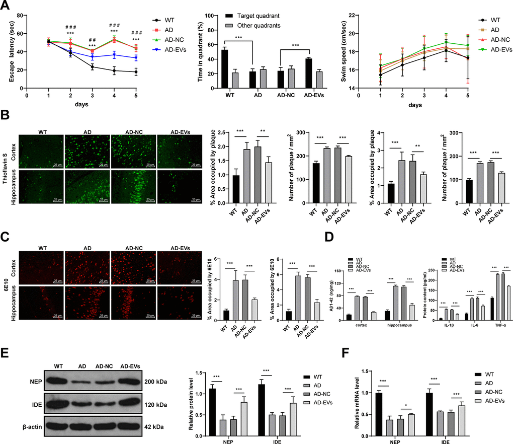
Figure 1.BM-MSC-EVs have therapeutic effects on AD rats. The rat model of AD was established by injection of Aβ1-42, and then rats were treated with BM-MSC-EVs, with injection of equal volume of BM-MSC conditioned medium after GW4869 treatment as the control. (A) Rat behaviors and memory abilities were measured using Morris Water maze test; (B) Thioflavin S staining for Aβ deposition in cerebral cortex and hippocampus of rats in each group; (C) Immunofluorescence assay was used to detect Aβ content in cerebral cortex and hippocampus of rats in each group; (D) ELISA was used to detect Aβ1-42 level in cerebral cortex and hippocampus and levels of inflammatory cytokines (IL-1β, IL-6 and TNF-α) in cerebral tissues of rats in each group; (E) WB was used to detect the protein levels of NEP and IDE; (F) RT-qPCR was used to detect the mRNA expression of NEP and IDE. n = 6. Data were expressed as mean ± standard deviation. Data were analyzed using one-way ANOVA followed by Tukey’s multiple comparisons test. In panel (A) ***p < 0.001 (WT vs AD), ### p < 0.001 (AD-NC vs AD-EVs). In other panels, *p < 0.05, **p < 0.01, ***p < 0.001.