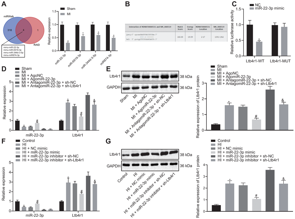
Figure 2.miR-22-3p could target Ltb4r1. (A) Prediction of miRNAs that bind to Ltb4r1 and RT-qPCR analysis of four candidate miRNA expression in MI model by miRWalk (http://mirwalk.umm.uni-heidelberg.de/) and RAID v2.0 (http://www.rna-society.org/raid/index.html). (B) The putative binding sites of miR-22-3p and Ltb4r1 3'UTR by the online website (http://bioinformatics.psb.ugent.be/webtools/venn/). (C) Luciferase activity of PGLO-Ltb4r1 WT and PGLO-Ltb4r1 MUT detected using dual-luciferase reporter gene assay upon treatment with NC and miR-22-3p mimic. (D) miR-22-3p expression and Ltb4r1 mRNA level in myocardial tissues of MI mice or sham-operated mice determined using RT-qPCR, upon treatment with AgomiR-22-3p, AntagomiR-22-3p, sh-Ltb4r1 or sh-NC. (E) Ltb4r1 protein level in myocardial tissues of MI mice determined using Western blot analysis upon treatment with AgomiR-22-3p, AntagomiR-22-3p, sh-Ltb4r1 or sh-NC. (F) miR-22-3p expression and Ltb4r1 mRNA level in hypoxia-induced MI cardiomyocytes determined using RT-qPCR. (G) Ltb4r1 protein level in hypoxia-induced MI cardiomyocytes determined using Western blot analysis, normalized to GAPDH. * p < 0.05 vs. hypoxia-induced cardiomyocytes treated with NC mimic or sham-operated mice or cardiomyocytes treated with empty vector, # p < 0.05 vs. MI mice injected with AgomiR NC or hypoxia-induced MI cardiomyocytes treated with NC mimic, and & p < 0.05 vs. MI mice injected with AntagomiR-22-3p + sh-NC or hypoxia-induced MI cardiomyocytes treated with miR-22-3p inhibitor + sh-NC. Unpaired t-test was used to analyze data between two groups and one-way ANOVA/Tukey’s test to analyzed data among multiple groups.