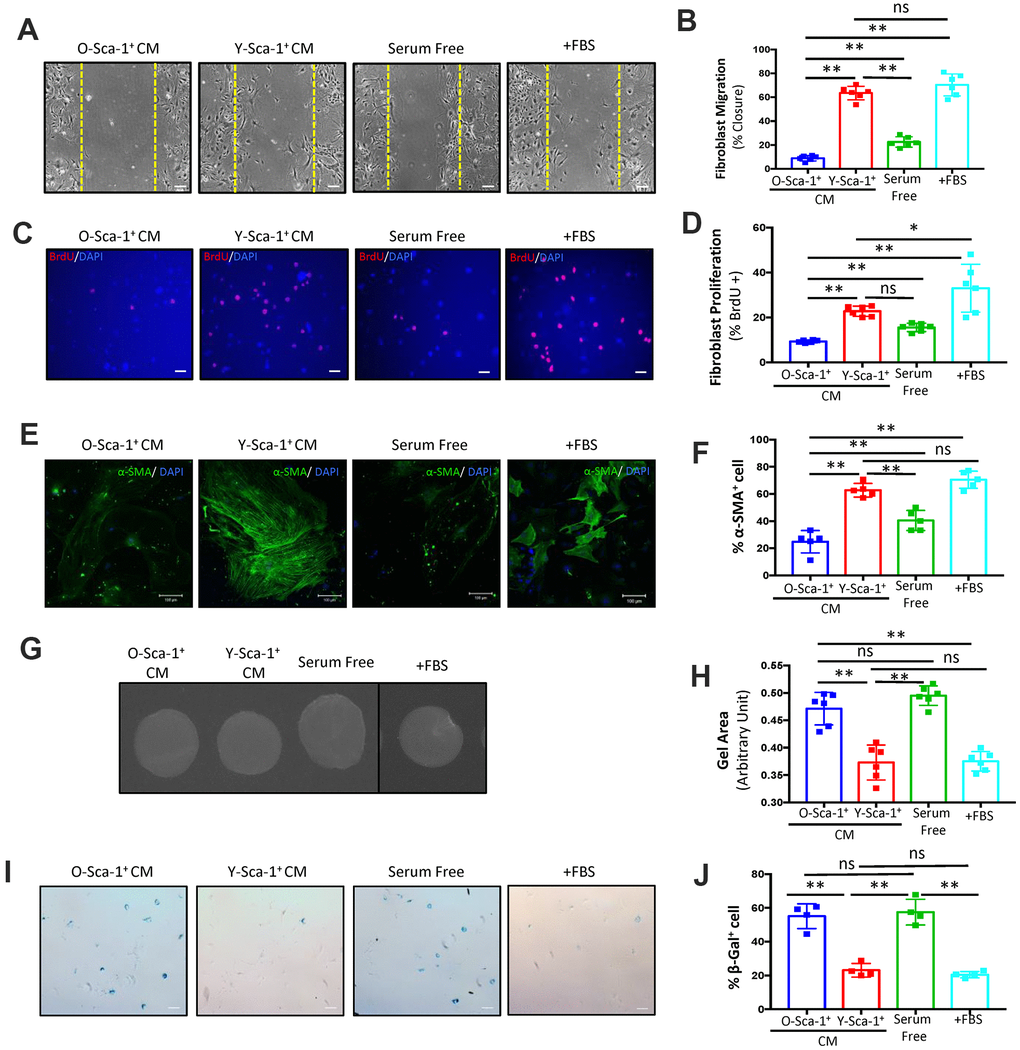
Figure 1.Conditioned media from Y-Sca-1+ BMCs improves functional and age-related deficits in old cardiac fibroblasts. (A) Representative images from scratch wound assay of old fibroblasts, treated with conditioned media (CM), from Y-Sca-1+ and O-Sca-1+ bone marrow cells (BMCs) for 48 hours. Dashed yellow line indicates the wound edge at 0 hours. After 48 hours, the closing distances were measured (B) (n=6). (C) Representative images from proliferation assay, after old fibroblasts were treated with CM from Y-Sca-1+ and O-Sca-1+ BMCs for 24 hours. BrdU is stained in red, and nuclei stained in blue. (D) Percentage of BrdU+ cells, normalized to total cell number. (E) Representative images of senescence assay (β-galactosidase+), after old fibroblasts were treated with CM from Y-Sca-1+ and O-Sca-1+ BMCs for 48 hours. (F) Percentage of SA-β-gal+ cells, normalized to total cell number (n=4). (G) Representative images of gels from gel contraction assay, after old fibroblasts were treated with CM from Y-Sca-1+ and O-Sca-1+ BMCs for 48 hours. (H) Gel area was measured using ImageJ (n=6). (I) Immunofluorescent staining for α-SMA was performed on old cardiac fibroblasts, after treatment with CM from Y-Sca-1+ and O-Sca-1+ BMCs for 48 hours. α-SMA is stained in green, and nuclei in blue. (J) Percentage of α-SMA+ cells, relative to total cell number (n=5). Scale bars represent 100 μm, unless otherwise stated. Data analysis was by one-way ANOVA. n=5-6; *p≤0.05, **p≤0.01; ns: not statistically significant.