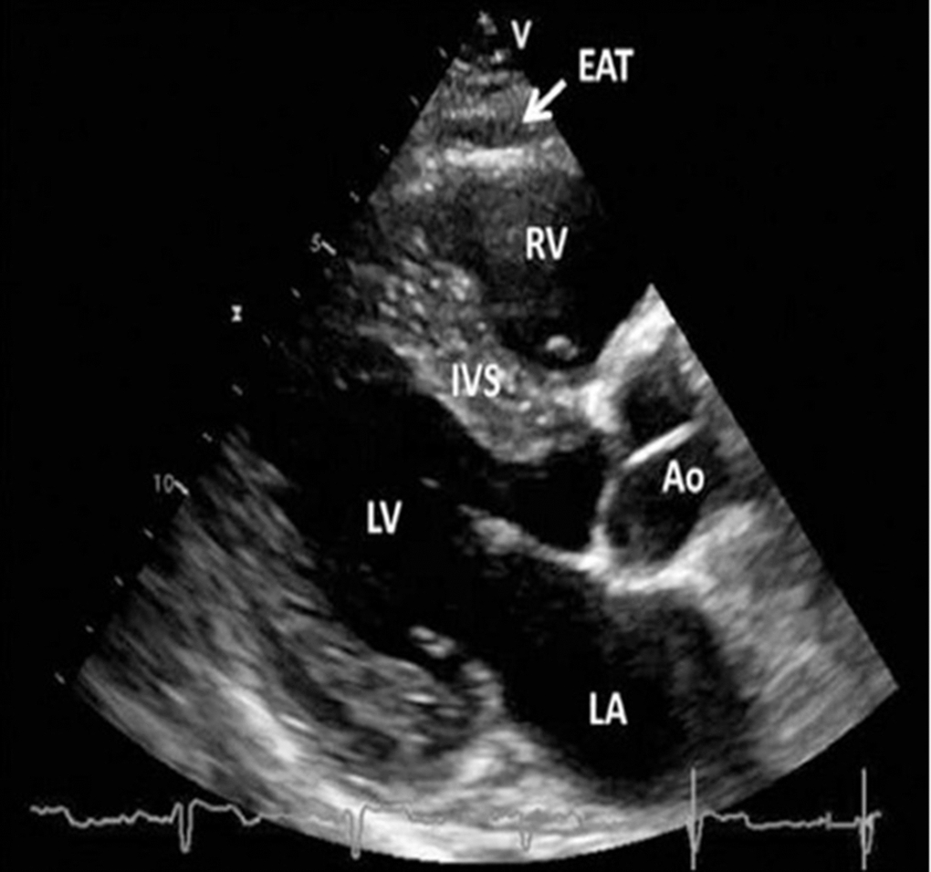
Figure 2.Transthoracic echocardiographic view of epicardial adipose tissue. Epicardial adipose tissue is an echo-lucent area between the epicardial surface and parietal pericardium in front of the right ventricular free wall and is indicated with a white arrow. Abbreviations: Ao, aorta; EAT, epicardial adipose tissue; IVS, interventricular septum; LA, left atrium; LV, left ventricle; RV, right ventricle [36, 37].