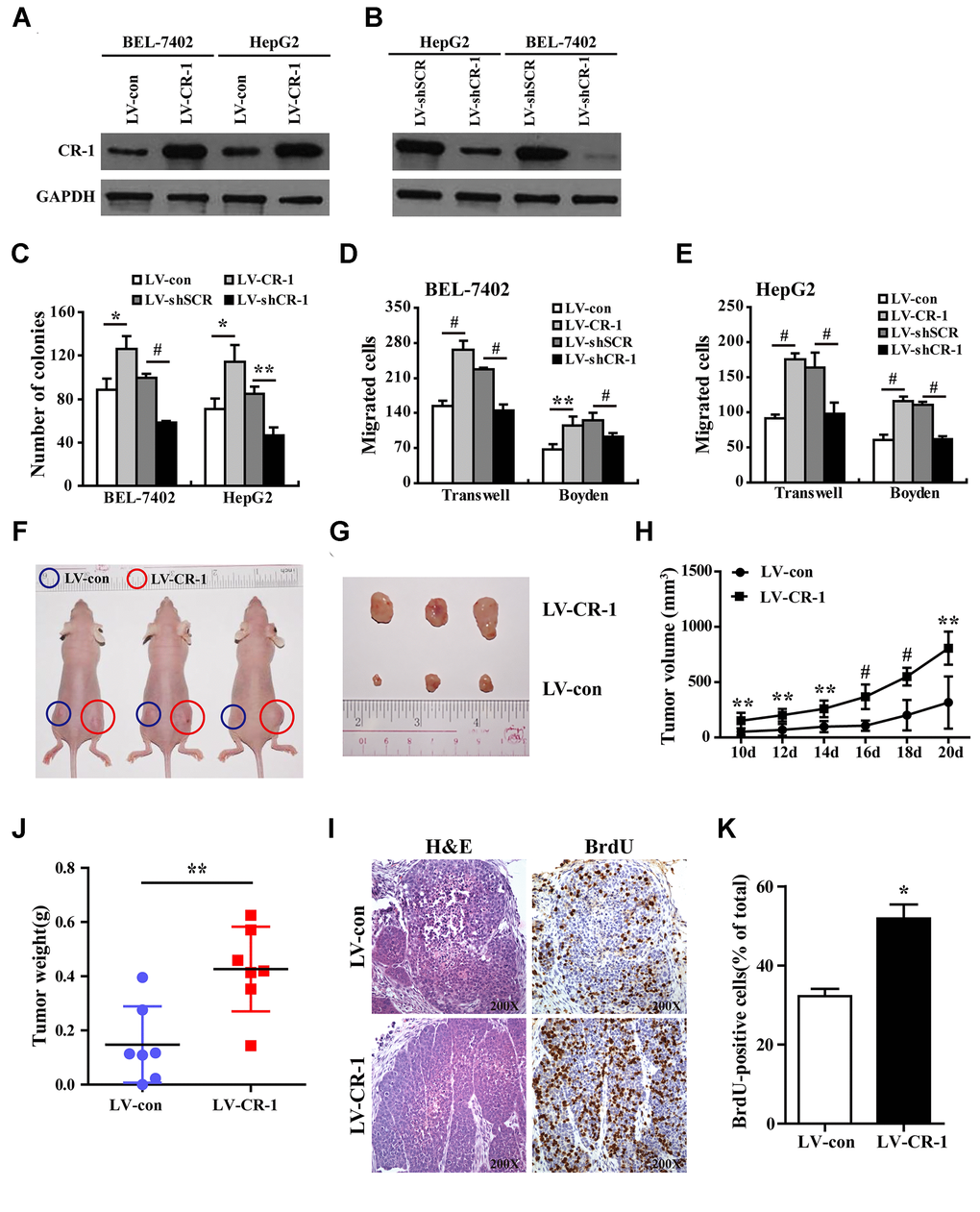
Figure 4.CR-1 overexpression promotes in vitro proliferation, migration and invasion of HCC cells and in vivo xenograft HCC growth in nude mice. (A) Western blot analysis shows CR-1 protein expression in control (LV-con) and CR-1-overexpressing (LV-CR-1) BEL-7402 and HepG2 cells. (B) Western blot analysis shows CR-1 protein expression in scrambled control (LV-shSCR) and CR-1-silenced (LV-shCR-1) BEL-7402 and HepG2 cells. (C) Colony formation assay results show proliferation ability of CR-1-overexpressing and CR-1-knockdown HCC cells with their corresponding controls. The representative images of this assay are shown in Supplementary Figure 5. (D–E) Transwell migration and Boyden invasion assay results show the migration and invasion abilities of CR-1-overexpressing, CR-1-knockdown, and control BEL-7402 (D) and HepG2 (E) cells, respectively. (F) Representative pictures of nude mice bearing subcutaneous xenografts from LV-con (blue circle) or LV-CR-1 (red circle) transduced BEL-7402 cells. (G) Representative images of subcutaneous xenograft tumors formed from LV-con or LV-CR-1 transduced BEL-7402 cells. (H) Growth curve of xenograft tumor volumes derived from LV-con or LV-CR-1 transduced BEL-7402 cells. (I) The weights of xenograft tumors derived from LV-con or LV-CR-1 transduced BEL-7402 cells. (J) Representative images of H&E-stained and BrdU-stained sections of xenograft tumors derived from LV-con or LV-CR-1 transduced BEL-7402 cells. (K) The percentages of BrdU-positive cancer cells in xenograft tumors derived from LV-con or LV-CR-1 transduced BEL-7402 cells, as calculated by total number of BrdU-positive cells relative to total number of cancer cells.