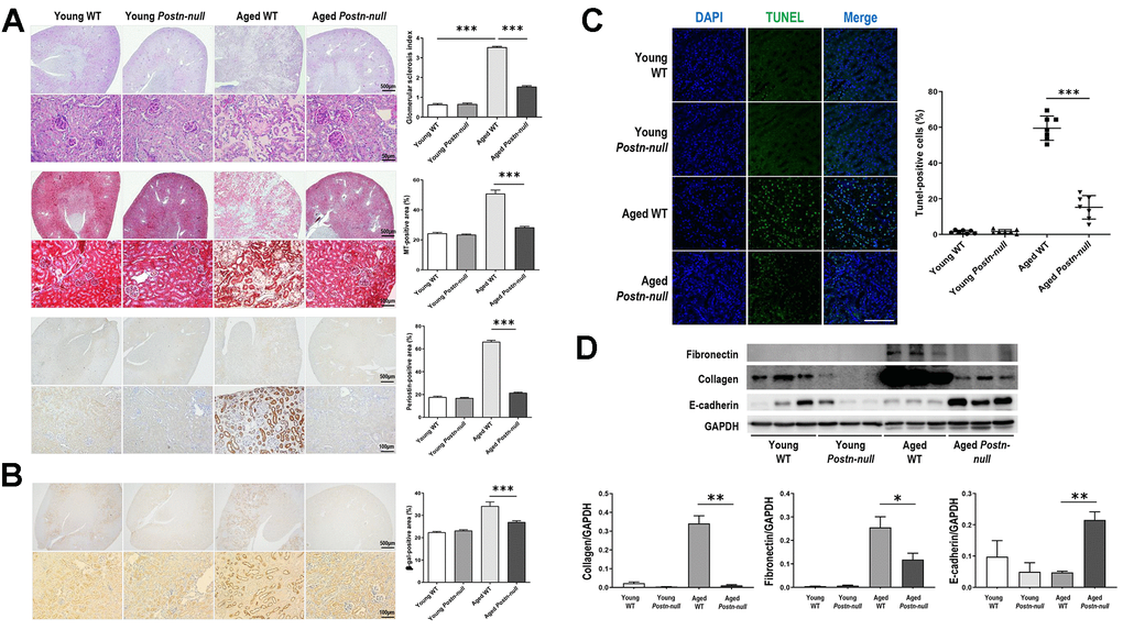
Figure 2.Role of periostin in renal fibrosis due to aging. (A) Tubular atrophy, interstitial fibrosis, glomerular sclerosis, and periostin expression were increased in aged WT mice, but not so much in aged Postn-null mice. Representative data are shown (N = 7/group). Magnification: 40× (top); 400× (bottom); 200× (bottom). Data are the mean ± SEM. ***p < 0.001 (unpaired t-test). (B) Beta-galactosidase expression was increased in aged WT mice, but a lesser extent in aged Postn-null mice. Representative data are shown (N = 7/group). Magnification: 40× (top); 200× (bottom). Data are the mean ± SEM. ***p < 0.001 (unpaired t-test). (C) Apoptotic cells were significantly increased in aged WT, but to a markedly lower level in aged Postn-null mice. Data are the mean ± SEM. ***p < 0.001 (unpaired t-test). (D) The expression of fibrosis markers was increased in aged WT mice, but not so much in aged Postn-null mice. Data are the mean ± SEM. *p < 0.05; **p < 0.01; ***p < 0.001 (unpaired t-test).