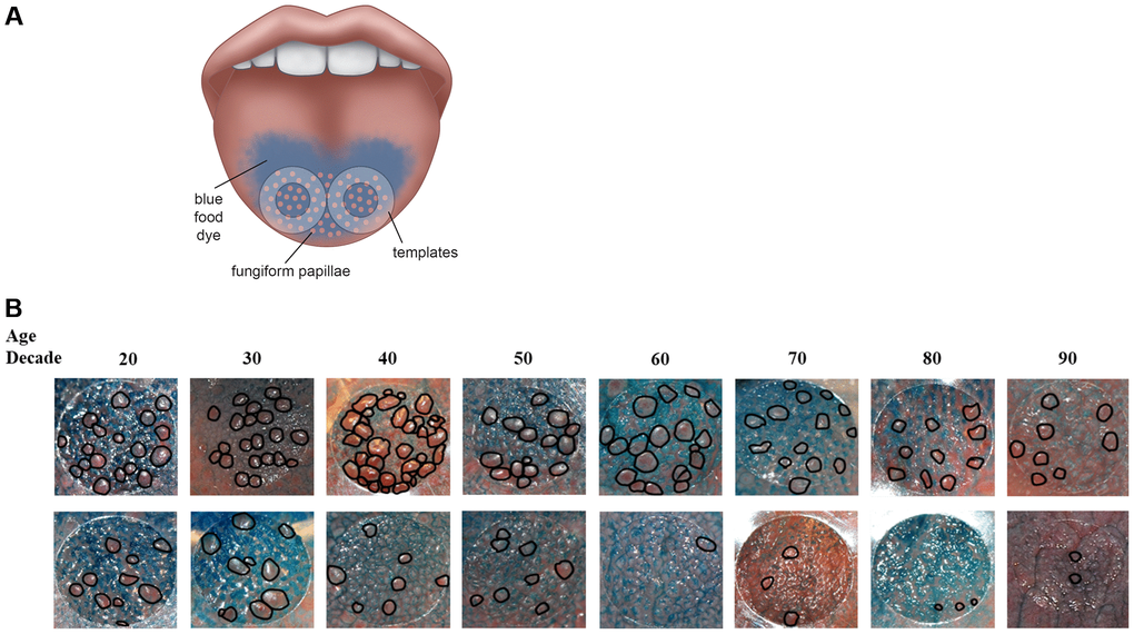
Figure 1.(A) Method to assess fungiform papillae density. Blue food coloring is used to provide optimal contrast between fungiform papillae (do not take up blue dye and appear pink) and other tongue structures (coated blue). Two clear plastic hole reinforcement templates (7 mm in diameter) are placed posterior to the apex of the tongue on each side of the median sulcus. Tongue images containing the two templates were taken using a digital camera. The fungiform papillae present within the two 7 mm holes were then counted and normalized to the area of the holes and expressed as fungiform papillae density (number of fungiform papillae/cm2). (B) Representative tongue images from 16 participants with age spans from the 20s to the 90s. As shown, fungiform papillae density varies widely among individuals and across lifespan. The top panel shows individuals with higher fungiform papillae density versus those individuals with lower fungiform papillae density in the lower panel.