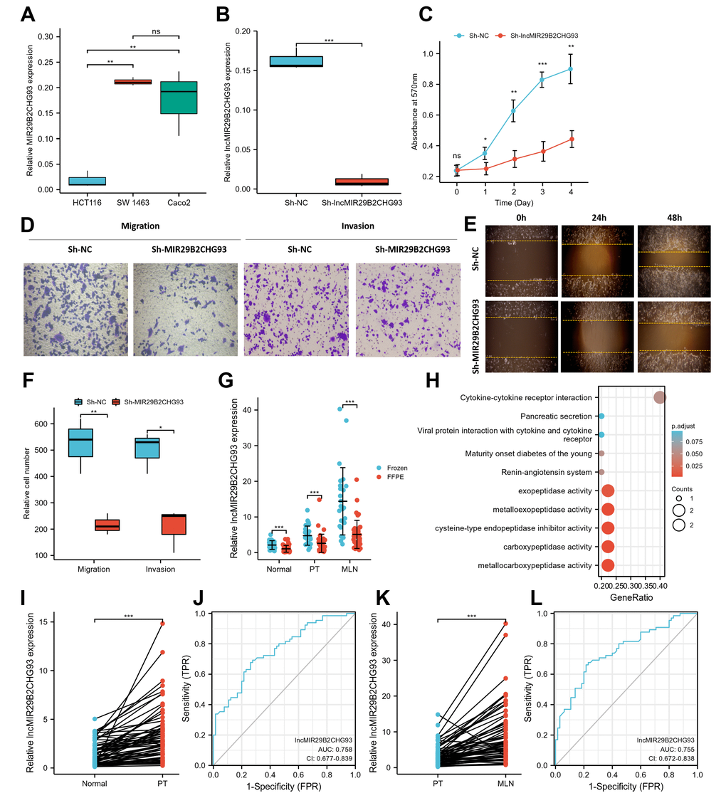
Figure 8.LncRNA MIR29B2CHG93 promoted cell proliferation and tumor metastasis in CRC. (A) RT-PCR analysis was used to detect the expression of lncRNA MIR29B2CHG93 in cell lines. (B) The expression levels of lncRNA MIR29B2CHG93 in Caco2 cells after transfection with sh-NC or sh-MIR29B2CHG93 were detected by RT-PCR. (C) The effects of lncRNA MIR29B2CHG93 knockdown on the proliferation of Caco2 cells were examined by MTT assay. (D–F) Transwell assay and wound healing assay were used to evaluate the migration and invasion ability of Caco2 cells transfected with sh-NC or sh-MIR29B2CHG93. (D) Images of Caco2 cells in migration and invasion transwell assays. (E) Cell mobility was determined by wound healing assay at 0, 24, 48h after the scratching. (F) Quantification of cell migration and invasion in (D). (G) The expression of lncRNA MIR29B2CHG93 was compared between in frozen tissue and FFPE tissues in normal mucosa, primary tumor and lymphnodal metastasis tumor tissue. (H) GO and KEGG analysis of lncRNA MIR29B2CHG93 based on co-expressed mRNAs. (I) LncRNA MIR29B2CHG93 expression in primary tumors compared with paired normal tissues from CRC cohort. (J) A ROC curve for assessing the predictive ability of lncRNA MIR29B2CHG in predicting normal and tumor. (K) LncRNA MIR29B2CHG93 expression in lymphnodal metastasis tumors compared with paired primary tumors from CRC cohort. (L) A ROC curve for assessing the predictive ability of lncRNA MIR29B2CHG in predicting lymphnodal metastasis. *P<0.05, **P<0.01, ***P<0.001.