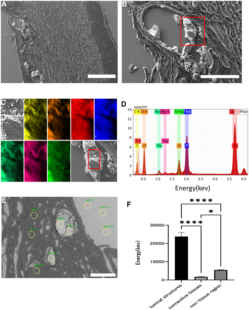
Figure 2.Scanning electron microscopy images and EDX analysis. Scanning electron micrograph of skin around an ulcer on a patient's elbow with WS (A: x 60, B: x 200). Crystalline substance in luminal structures under the dermis (red squares). This crystalline substance was examined with energy dispersive X-ray (EDX) (C, D). Each color and element combination corresponds to C for yellow, O for orange, Na for light blue, Mg for pink, Si for green, P for blue, and Ca for red. Using EDX, we measured the calcium content of the luminal structure, dermal connective tissue, and non-tissue areas at three points in each area (yellow circles), and performed similar measurements in three fields (E). Comparison of the calcium content of the luminal structure, dermal connective tissue, and non-tissue areas (F). Data are expressed as mean ± standard error. One-way ANOVA followed by Tukey test were performed (* p < 0.01, **** p < 0.0001).