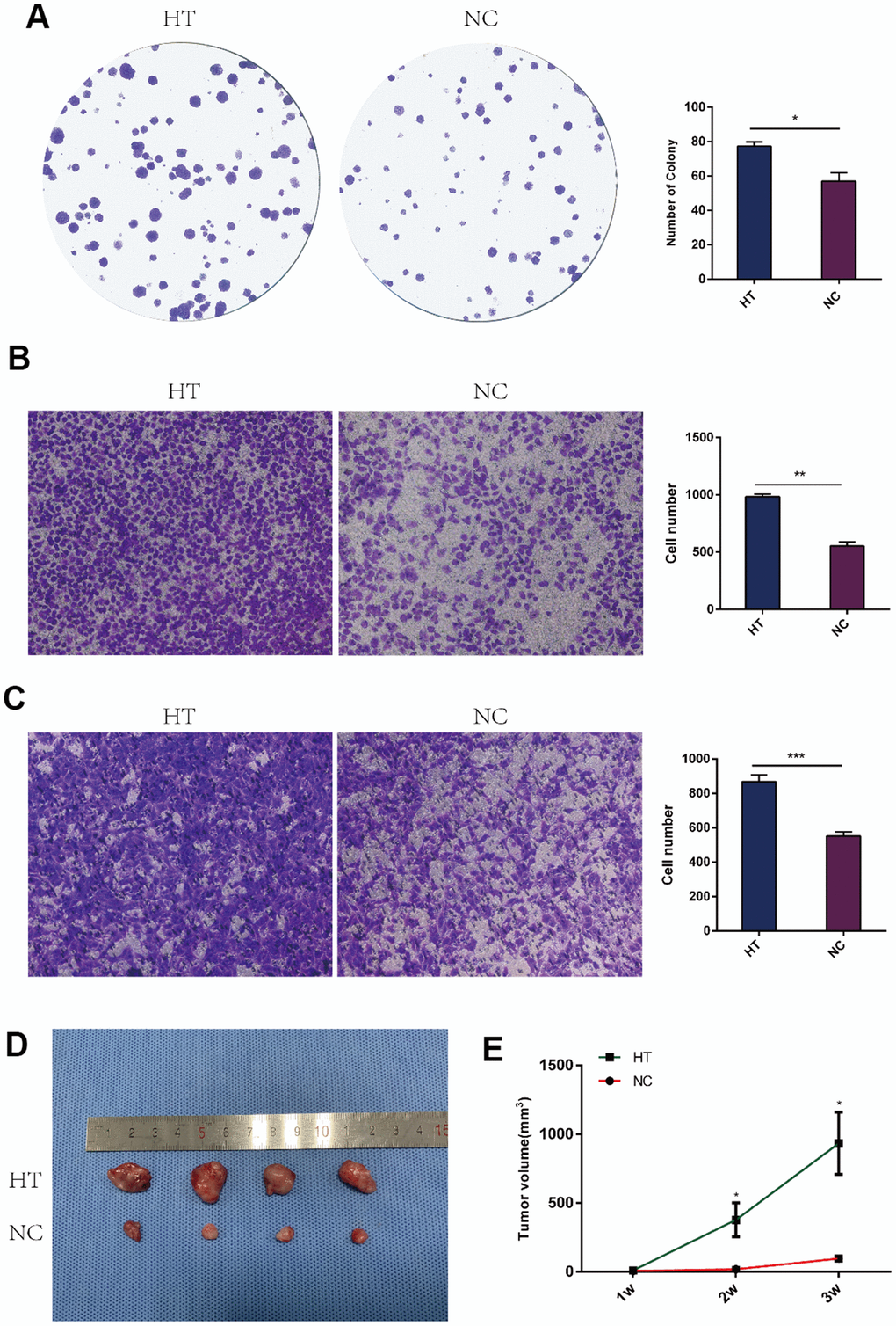
Figure 1.Sublethal heat treatment promoted the proliferation and invasion of 4T1 cells. (A) Colony formation assays were performed to assess the proliferation of 4T1 cells after sublethal heat treatment. The colonies were identified and counted. The number of colonies were presented as histograms. (B) Transwell assays were performed to determine the invasive ability of 4T1 cells after sublethal heat treatment. Representative images of invasive cells in the lower chamber stained with crystal violet. (C) Transwell assays were performed to determine the invasive ability of HUVECs after co-cultured with supernatant of sublethal heat treated 4T1 cells. The quantification of cell invasion was presented as invaded cell numbers. (D)The tumor grafts were showed (n=4 in each group) at the end of the 3rd week; (E) Tumor volumes were recorded and compared every week. All data were expressed as mean±SD of three independent experiments. HT=high temperature (45° C), NC=negative control (37° C). * indicates P<0.05, ** indicates P<0.01, *** indicates P<0.001.