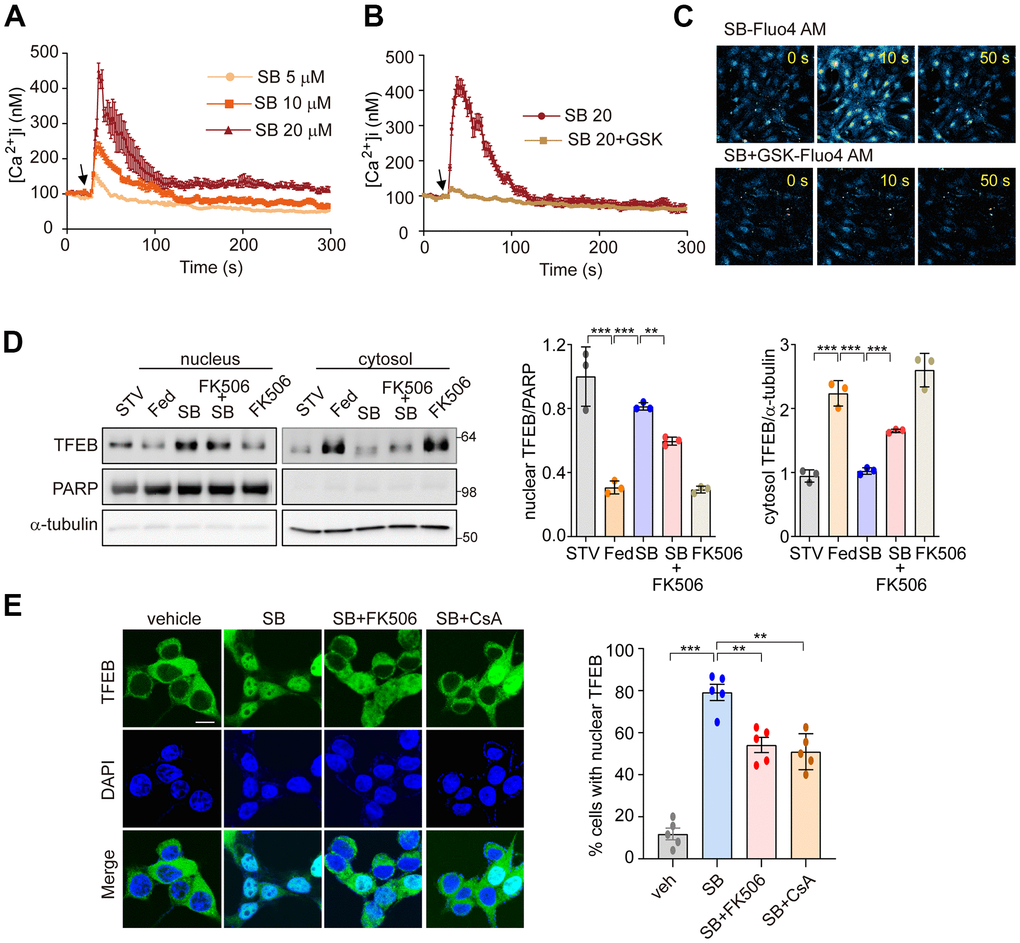
Figure 4.PERK-Ca2+-calcineurin pathway is required for SB202190-induced TFEB nuclear translocation. (A–C) The change of [Ca2+] in MEF cells was measured using confocal microscopy after loading with Fluo-4 AM. (A) Arrow indicates the time point at which SB202190 (SB) was added. The data represent mean ± SD from 3 independent experiments. (B, C) MEF cells were preincubated for 30 min with the PERK inhibitor GSK2606414 (1 μM). Arrow indicates the time point at which 20 μM SB202190 was added. The data represent mean ± SD from 3 independent experiments. (D) SH-SY5Y cells were pretreated with FK506 (10 μM) for 30 min and then treated with SB202190 (20 μM) for another 3 h. Measurement of TFEB activation was performed by western blotting of nuclear and cytoplasmic extracts. TFEB expression in the nucleus and cytoplasm was normalized to PARP and α-tubulin, respectively (left). Quantification of TFEB translocation is shown in the right panel. Data represent mean ± SD, **p<0.01; ***p<0.001. (E) TFEB-GFP-transfected SH-SY5Y cells were treated with SB202190 (20 μM) for 6 h in the presence or absence of calcineurin inhibitors, FK506 (10 μM) and Cyclosporin A (CsA, 20μM). Representative images were detected by confocal microscopy (left). Quantification of nuclear translocation of TFEB-GFP (right). n > 20 cells per condition. Data represent mean ± SD; **p<0.01 and ***p<0.001.