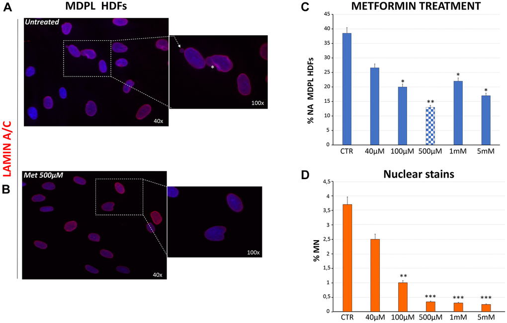
Figure 5.Evaluation of nuclear shape organization after metformin treatment at different concentrations. (A) Representative image of nuclear shape organization observed in MDPL HDFs stained for lamin A/C (red), showing the presence of membrane invaginations (asterisks), and micronuclei (white arrows). (B) Representative image of lamin A/C immunostaining in MDPL HDFs following 48 h of treatment with 500 μM of metformin, in which a clear reduction in nuclear anomalies is evident. (C) Evaluation of aberrant nuclear alteration (% NA) in MDPL HDFs treated for 48 h with increasing doses of metformin. Each value represents the mean ± SD. of the analysis of 300 cells observed in three independent experiments (*p < 0.05; **p < 0.01). Values are displayed as the average percentages of two different patients (D) Percentage of micronuclei (MN) encountered in MDPL-HDFs after 48 h of metformin treatment. The data have been obtained counting the micronuclei after Hoechst 33342 nuclear staining for fluorescence imaging. Each value represents the mean ± S.D. of the analysis of 300 cells for three independent experiments (**p-value <0.01; ***p-value <0.001). Values are displayed as the average percentages of two different patients. Hoechst 33342 nuclear staining (blue). Magnification 40× and 100×. Abbreviations: HDFs: human dermal fibroblasts; Met: metformin; MN: micronuclei; NA: aberrant nuclear alteration; Untreat: untreated cells.