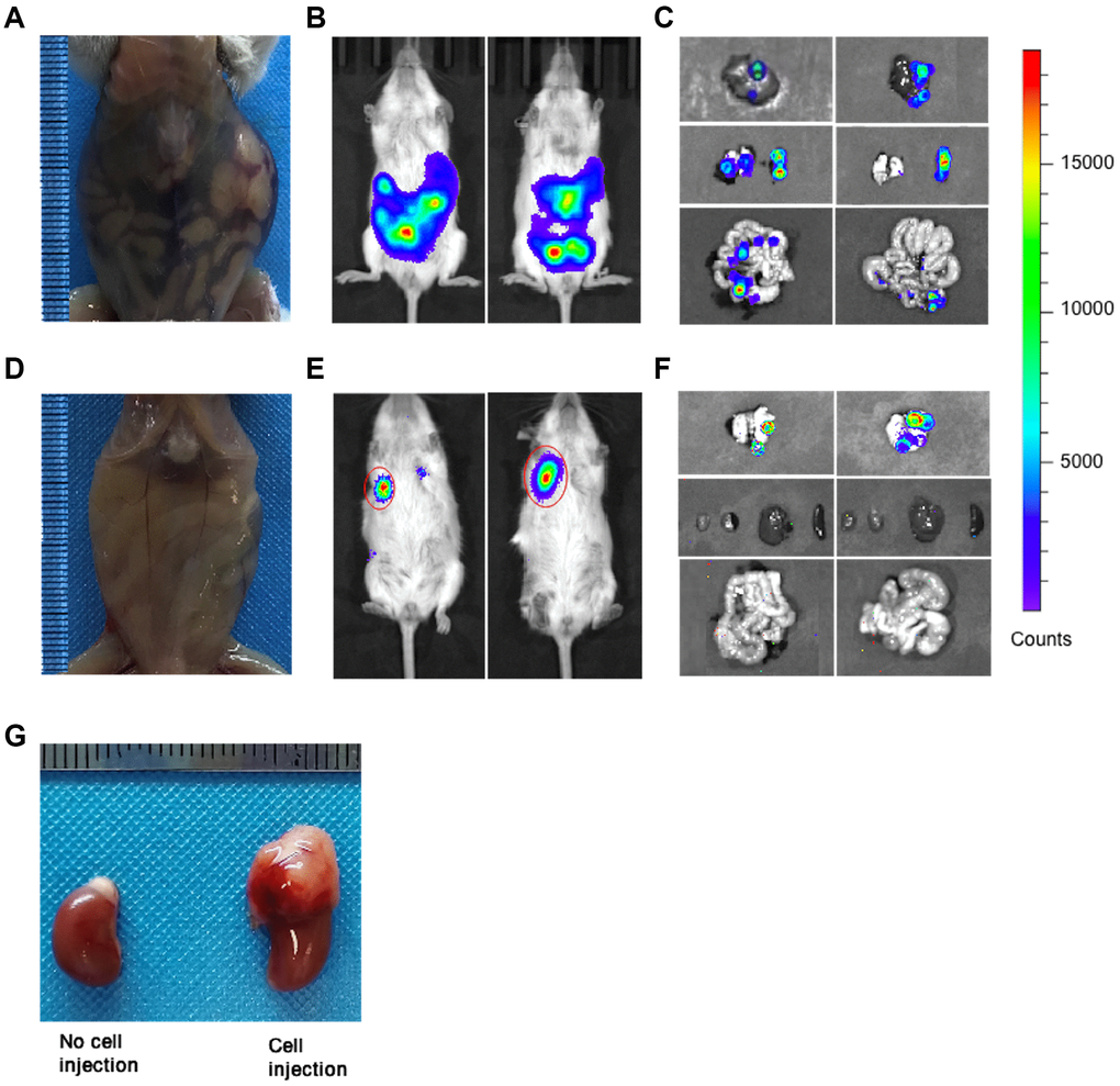
Figure 2.Tumor metastasis in orthotopic and subcutaneous CDX mice. (A) Representative image of orthotopic CDX mice was taken at day 14 after cell implantation (Mice used, n = 6, Representative data used, n = 1). (B) Representative bioluminescence images of tumors in orthotopic CDX mice (Mice used, n = 6, Representative data used, n = 2). (C) Representative bioluminescence images for the metastatic liver, lung, spleen and intestine in orthotopic CDX mice (Mice used, n = 6, Representative data used, n = 2). (D) Representative image of subcutaneous CDX mice were taken at day 21 after cell implantation (Mice used, n = 5, Representative data used, n = 1). (E) Representative bioluminescence images of tumors in subcutaneous CDX mice (Mice used, n = 5, Representative data used, n = 2). (F) Representative bioluminescence images for the metastatic liver, lung, spleen and intestine in subcutaneous CDX mice (Mice used, n = 5, Representative data used, n = 2). (G) Representative images for the kidneys and the adrenal glands were injected with or without cells (two kidneys from one CDX mouse). Left: Kidney without cell injection. Right: Kidney with cell injection.