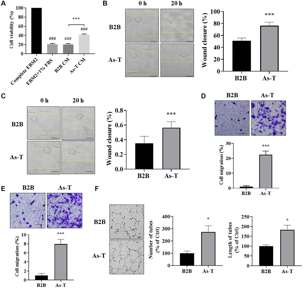
Figure 3.Vascular endothelial cells cultured in conditioned medium (CM) from As-T cells showed higher cell viability, more cell migration, and better tube formation. (A) HUVECs were incubated with the CM from B2B or As-T cells for 4 days. Cell proliferation of HUVECs was determined by counting the number of trypan blue negative cells. ###p < 0.001 compared to complete EBM2 medium group; ***p < 0.001 compared to B2B CM group. (B) When reached about 100% confluence, HUVECs were starved overnight, and a scratch wound was made as above. Superannuant was removed, and the CM from B2B or As-T cells was added to the wells. The widths of the wounds were measured at 0 h and 20 h post scratch. Images were taken using an inverted microscope at 10× magnification, bar = 100 μm. Left panel: representative images; right panel: quantification of the wound healing assay. ***p < 0.001 compared to CM from the B2B group. (C) Wound healing assay for the HMVECs was performed as described above. Bar = 100 μm, ***p < 0.001. (D) A transwell assay was conducted to evaluate the effects of CM on HUVEC migratory ability. HUVECs were plated on the top chamber in 200 μL basic EBM2 medium; the lower chamber was filled with 600 μL CM from B2B or As-T cells. The cells that pass through the network were stained with crystal violet and counted under a microscope at 24 h, and the images were taken using a microscope at 10× magnification, bar = 100 μm. Upper panel: representative images; lower panel: quantification of the migrated cells. ***p < 0.001 compared to CM from B2B cells. (E) Migration assay for the HMVECs was performed as described above. Bar = 100 μm, ***p < 0.001. (F) A tube formation assay was carried out to evaluate the effects of CM on the tube forming ability of HUVECs. HUVECs were starved with basic EBM2 medium overnight and resuspended in 100 μL diluted CM (CM: basic EBM2 = 1:1). The cell suspension was then added to a 96 well-plate containing 100 μL solidified growth factor-reduced Matrigel. The tubular structures were imaged at about 6–12 h under a microscope at 4× magnification. Left panel: representative images of the tubular structures. Middle panel: quantification of the number of the tubular structures. Right panel: quantification of the length of the tubular structures. *p < 0.05 compared to CM from B2B group.