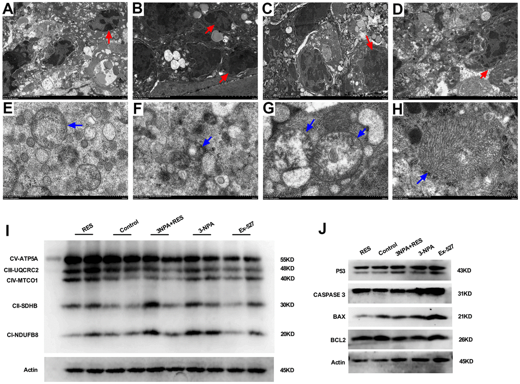
Figure 6.Mitochondrial oxidative phosphorylation-induced apoptosis may result in oxidative stress-induced POI. (A) Granulosa cells in ovarian tissue from the control group. (B) In the 3-NPA model group, GCs apoptosis and nuclear pyknosis were obvious, the chromatin margin was set, organelle structure was destroyed, and empty shots appeared. (C) Obvious granulocyte apoptosis was observed in ovarian tissue from mice in the Ex-527 intervention group; chromatin agglutination and vacuoles were also observed. (D) Ovarian granulocyte apoptosis was significantly lower in the resveratrol intervention group than in the model group. (E) Mitochondrial morphology in ovarian tissue from mice in the control group was largely normal, and the number of mitochondria was high. (F) In the 3-NPA group, mitochondrial morphology in ovarian tissue was destroyed, stromal edema was obvious, and the endoplasmic reticulum was dilated and damaged. (G) Ovarian mitochondria in mice from the Ex-527 intervention group were obviously swollen and cavitated. (H) Ovarian mitochondria in the resveratrol intervention group showed significantly improved morphology; the mitochondrial crest was clearly visible, and the mitochondrial matrix was uniform. (I) OXPHOS expression was down-regulated in the Ex-527 and 3-NPA groups, while it was up-regulated in the Res group. (J) P53, BAX, and CASPASE3 were up-regulated in the Ex-527 and 3-NPA groups and down-regulated in the Res group. BCL2 was down-regulated in the Ex-527 and 3-NPA groups, while it was up-regulated in the Res group. Red arrow: granular nucleus; Blue arrow: mitochondria.