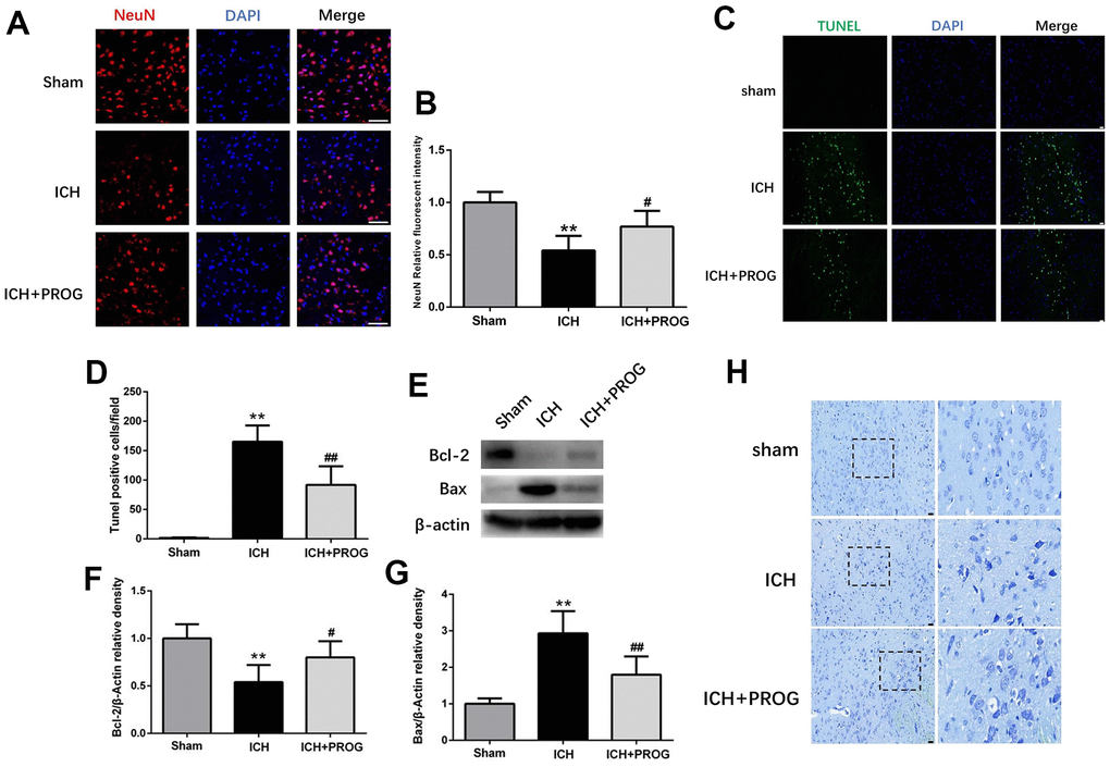
Figure 2.Effect of progesterone on neuron death and cell apoptosis. (A) Representative immunofluorescence staining images of NeuN (red) in perihematomal region. Nuclei were counterstained with DAPI (blue). Bar=50μm. (B) Quantitative analyses of NeuN relative fluorescent intensity in perihematomal region in each group. (C) Representative micrographs of TUNEL staining in perihematomal region in each group. Bar=50μm. (D) Quantitative analysis shows that TUNEL positive cells in perihematomal region. (E) Representative Western bands showing the protein expression of Bcl-2 and Bax in perihematomal region. (F, G) Quantitative analysis of Western blots shows that the expression of Bcl-2 and Bax changes in each group. (H) Representative Nissl-stained images in perihematomal region in each group. Bar=50μm. n = 6 animals per group. Data are expressed as the mean ± SEM; **P < 0.01 vs. sham; #P < 0.05 vs. ICH group; ##P < 0.01 vs. ICH group. PROG: progesterone.