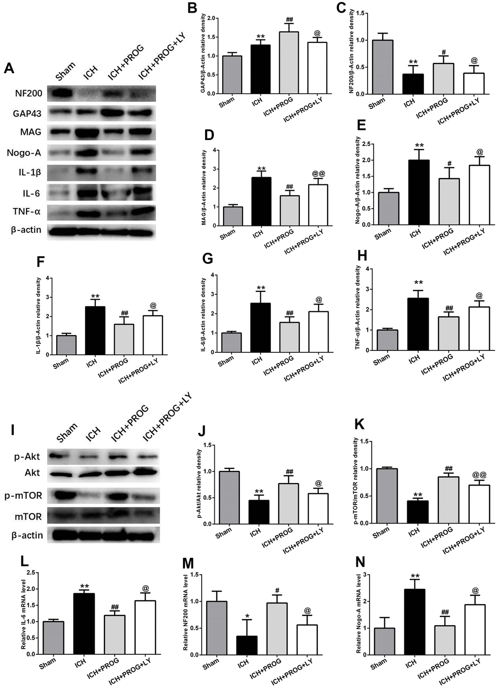
Figure 5.The mechanism of the effects of progesterone on anti-inflammation and promoting axonal regeneration. (A) Representative Western bands showing the protein expression of NF200, GAP43, Nogo-A, MAG IL-1β, IL-6 and TNF-α in perihematomal region.(B–H) Quantitative analysis of Western blots shows that the expression of NF200, GAP43, Nogo-A, MAG IL-1β, IL-6 and TNF-α changes in each group. (I) Representative Western bands showing the protein expression of p-Akt, total-Akt, p-mTOR and total-mTOR in perihematomal region. n = 6 animals per group. (J, K) Quantitative analysis of Western blots shows that the expression of p-Akt/total-Akt, p-mTOR/total-mTOR changes in each group. (L–N) mRNA expression levels of inflammatory and axon-related markers. n = 3 animals per group. Data are expressed as the mean ± SEM; *P < 0.05 vs. sham; **P < 0.01 vs. sham; #P < 0.05 vs. ICH group; ##P < 0.01 vs. ICH group; @ P < 0.05 vs. ICH+ progesterone group; @@ P < 0.01 vs. ICH+ progesterone group. LY:LY294002.