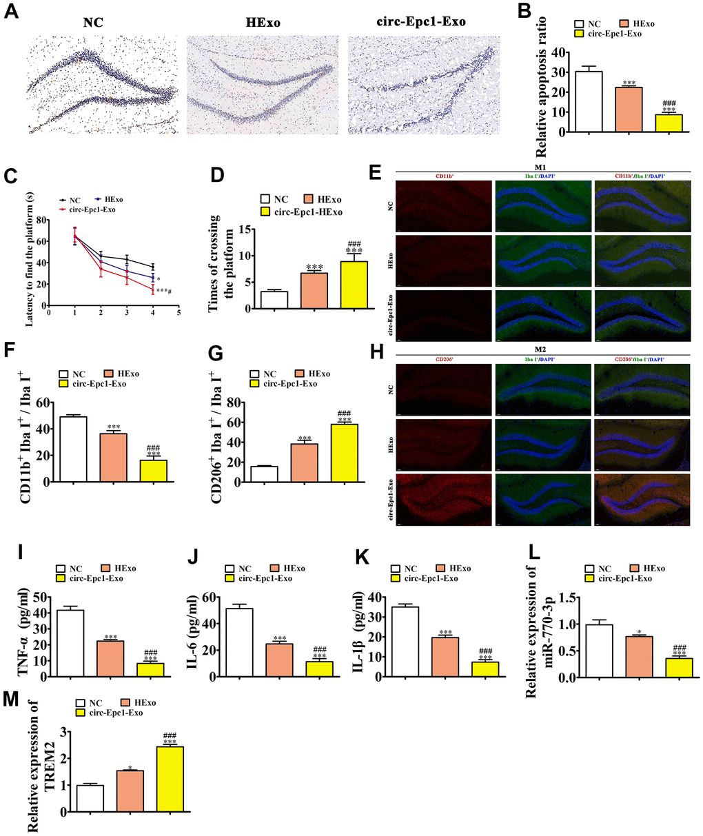
Figure 6.Circ-Epc1-modified ADSC exosomes (circ-Epc1-Exo) showed increased therapeutic effects at improving cognitive function by decreasing neuronal damage and shifting hippocampal microglia from M1 to M2. (A, B) Hippocampal neuron apoptosis was detected using the TUNEL assay. Data represent the mean ± SD (n=6). **P < 0.01, ***P < 0.001 vs. the control; #P < 0.05 vs. Exo. (C) Alzheimer’s disease mice exhibited a longer escape latency than exosome-treated animals. Data represent the mean ± SD (n=10). *P < 0.05, **P < 0.01 vs. the control; #P < 0.05 vs. hypoxia-pretreated ADSC exosomes (HExo). (D) The number of platform crossings was increased in the exosome-treated group. Data represent the mean ± SD (n=10). *P < 0.05, ***P < 0.001 vs. the control; ###P < 0.001 vs. HExo. (E–H) Immunofluorescence detection of macrophage polarization using F4/80+, CD11b+, and CD206+staining. Data are presented as the mean ± SEM. ***P < 0.001 vs. control; ###P < 0.001 vs. HExo. (I–K) ELISA results showing expressions of the inflammatory factors, TNF-α, IL-6, and IL-1β. Data are presented as the mean ± SEM. ***P < 0.001 vs. the control; ###P < 0.001 vs. HExo. (L, M) RT-qPCR showing the expressions of miR-770-3p and TREM2 in hippocampal tissues. Data are presented as the mean ± SEM. ***P < 0.001 vs. the control; ###P < 0.001 vs. HExo.