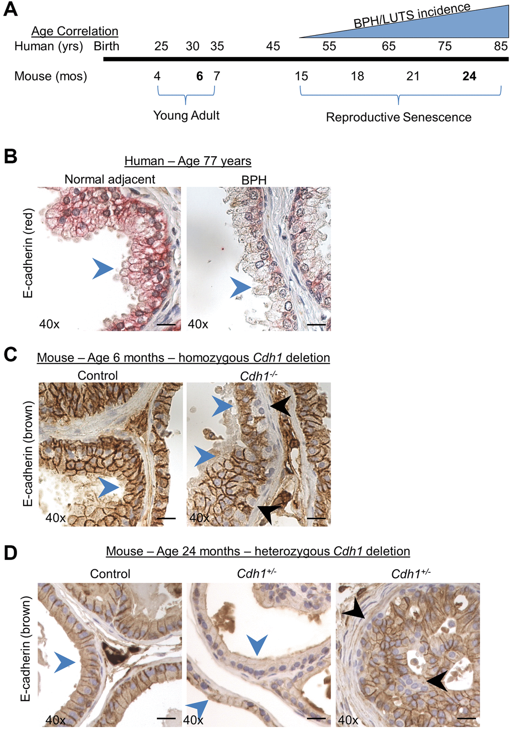
Figure 1.Modeling the combined impact of heterozygous E-cadherin loss and aging on the murine prostate. (A) Correlation of human age and BPH/LUTS incidence to aging of C57BL/6 mice [18]. Experimental timepoints for mice at 6 months and 24 months of age (noted in bold) correlate to 30 years and 75–85 years of age for men. (B) Representative E-cadherin immunostaining (red) in normal prostate adjacent to BPH (left) and in glandular BPH (right) specimens from a 77-year-old patient. (C) E-cadherin immunostaining (brown) in Control (left) and mice with homozygous deletion of E-cadherin (Cdh1-/-) (right) mouse ventral prostate at 6 months of age. E-cadherin immunostaining in Cdh1-/- mice displayed a mosaic pattern of E-cadherin negative cells (black arrows) surrounded by a layer of E-cadherin positive epithelial cells (blue arrows). (D) E-cadherin immunostaining in Control (left) and mice with heterozygous deletion of E-cadherin (Cdh1+/-) mouse ventral prostate (center showing apparent reduced E-cadherin immunostaining, and right panel showing mosaic staining pattern) at 24 months of age. Blue arrows indicate epithelial cells with positive E-cadherin staining, black arrow indicates E-cadherin negative epithelial cells. Original magnification, 40×. Scale bars indicate 25 μm.