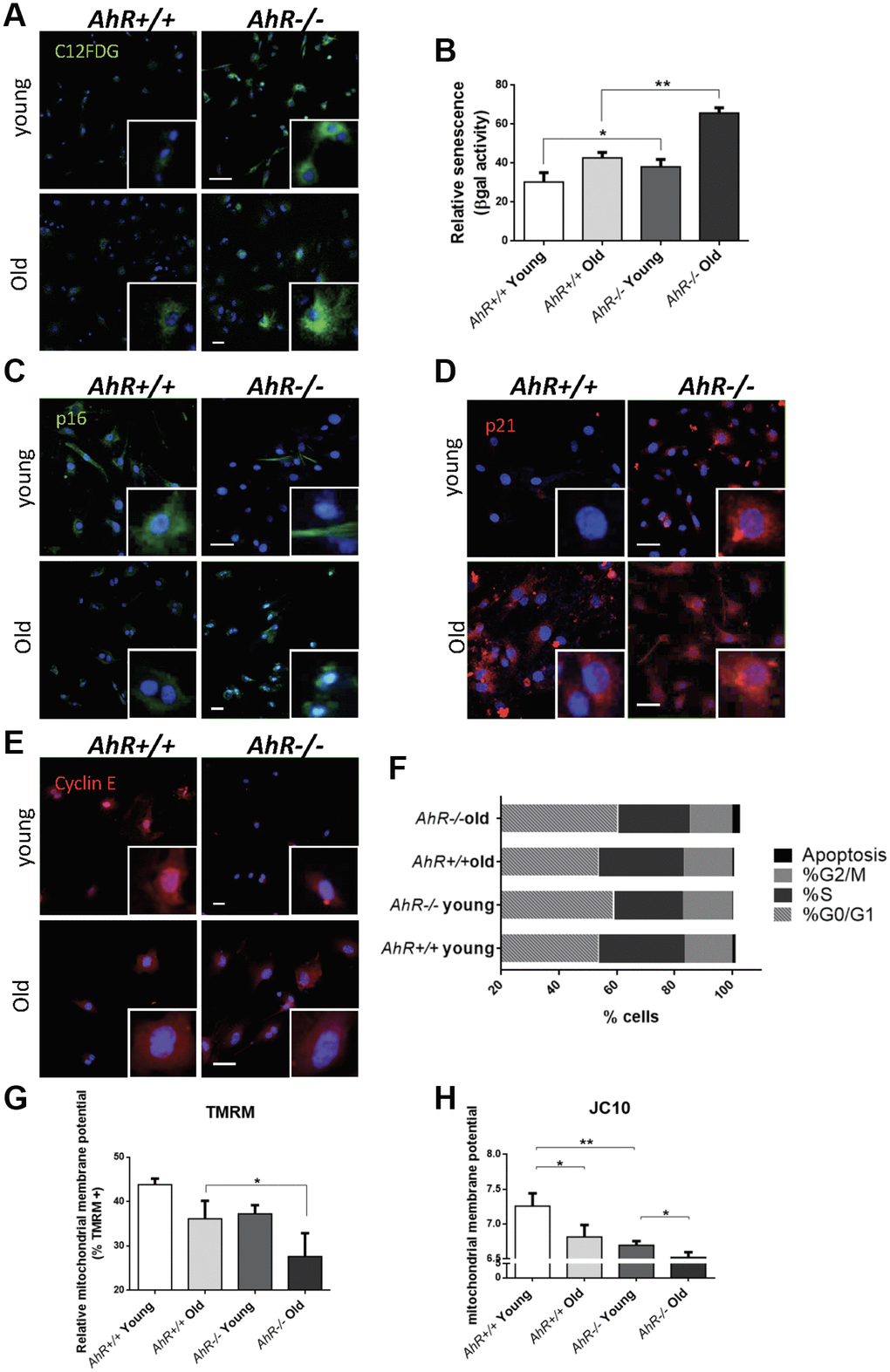
Figure 5.Senescence increases with aging in adult AhR-null fibroblasts. (A) AhR+/+ and AhR−/− fibroblasts were stained with the SA-β-Gal fluorescent substrate C12FDG to determine senescence levels by confocal microscopy. (B) SA-β-Gal activity was also measured by flow cytometry analyzing the percentage of C12FDG positive cells. (C–E) p16Ink4a (C), p21Cip1 (D) and Cyclin E (E) were analyzed by florescence confocal microscopy using specific antibodies in young and aged fibroblast cells. Conjugated secondary antibodies used were Alexa 488 and Alexa 633. DAPI staining was used to label cell nuclei. An Olympus FV1000 confocal microscope with FV10 software (Olympus) were used. (F) Percentage in each cell cycle phase. Cell cycle analysis was performed by FACS using propidium iodide staining. (G) Mitochondrial membrane potential (MMP) was quantified by the percentage of TMRM positive cells analyzed by Cytoflex S cytometer (Beckman Coulter). (H) MMP was also calculated using the JC10 kit for mitochondrial membrane polarization (Sigma-Aldrich). The red/green fluorescence intensity ratio was used to determine MMP activity. Data are shown as mean + SD (*P < 0.05; **P < 0.01).