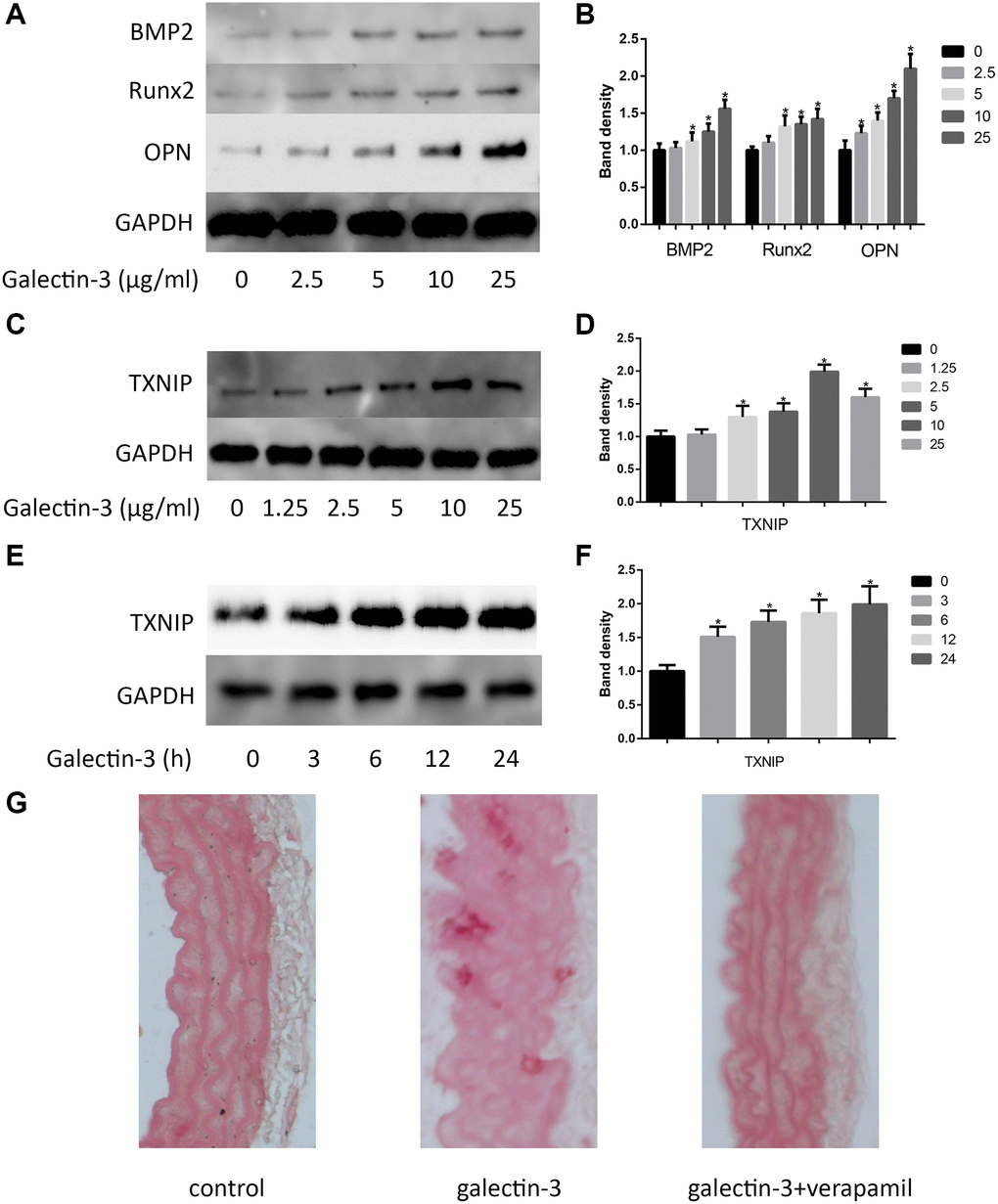
Figure 1.Galectin-3 induced aorta and VSMCs calcification. Cells were treated with galectin-3 over a range of concentrations (0, 2.5 μg/ml, 5 μg/ml, 10 μg/ml, 25 μg/ml) for 24 h, VSMCs osteogenic differentiation proteins (BMP2, Runx2 and OPN) were measured by Western blot, the quantification result is shown in the right panel (A and B). VSMCs were treated with galectin-3 over a range of concentrations (0, 1.25 μg/ml, 2.5 μg/ml, 5 μg/ml, 10 μg/ml, 25 μg/ml) for 24 h, TXNIP expression was measured by Western blot (C), the quantification result is shown in the right panel (D). 10 μg/ml galectin-3 was used to deal with VSMCs for different times (0, 3 h, 6 h, 12 h, 24 h), TXNIP expression was measured by Western blot (E), the quantification result is shown in the right panel (F). Band density of native VSMCs was defined as a control and considered to 1. Representative images of Alizarin Red staining of aorta (G). All experiments were performed at least three times. *P < 0.05 compared with control.