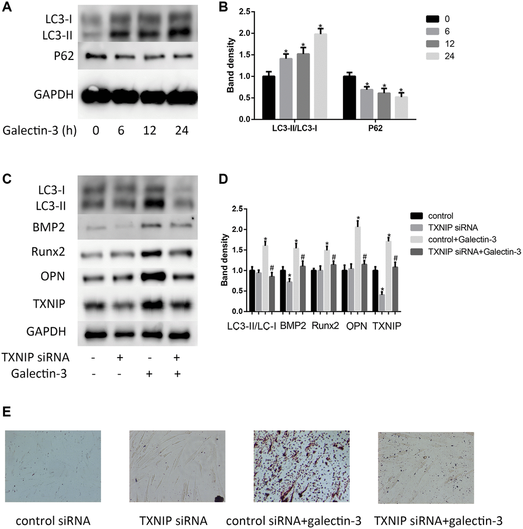
Figure 2.Galectin-3 induced VSMCs calcification and autophagy via TXNIP. VSMCs were treated with 10 μg/ml galectin-3 for different times (0, 6 h, 12 h, 24 h). The protein expression level of LC3-II/LC3-I and P62 were measured by western blot, the results quantifications were shown in the right panel (A and B). After transfection with either control or TXNIP siRNA for 24 h, VSMCs were incubated in the absence or presence of 10 μg/ml galectin-3 for 24 h, the expression of LC3-II/LC3-I and VSMCs osteogenic differentiation proteins (BMP2, Runx2 and OPN) was measured by western blot, the results quantifications were shown in the right panel (C and D). Band density of native VSMCs was defined as a control and considered to 1. After transfection with either control or TXNIP siRNA for 24 h, VSMCs were incubated in the absence or presence of 10μg/ml galectin-3 for 7 d, Alizarin red staining was used to observe the calcium deposition (E). Data were obtained from three independent experiments. *P < 0.05 vs. control; #P < 0.05 vs. galectin-3.