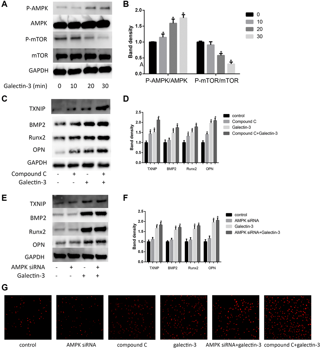
Figure 5.AMPK signaling pathway mediated galectin-3-induced TXNIP and VSMCs osteogenic differentiation. Cells were treated with 10 μg/ml galectin-3 over a range of times (0–60 min), and the expression of AMPK, p-AMPK, mTOR, and p-mTOR were measured by Western blot. Western blot results are shown, quantification of the results is given in the right panel (A and B). Band density of native VSMCs was defined as a control and considered to 1. Data were obtained from three independent experiments. *P < 0.05 compared with control. After pre-treatment with 1 μmol/L compound C for 1 h, VSMCs were then treated with 10 μg/ml galectin-3 for 24 h, the expression of TXNIP and VSMCs osteogenic differentiation proteins was measured by western blot (C). Quantification of the results were shown in the right panel (D). After pre-treatment with AMPK siRNA for 24 h, VSMCs were then treated with 10 μg/ml galectin-3 for 24 h, the expression of TXNIP and VSMCs osteogenic differentiation proteins was measured by western blot (E). Quantification of the results were shown in the right panel (F). Band density of native VSMCs was defined as a control and considered to 1. DHE staining was used to observe the ROS production (G). Data were obtained from three independent experiments. *P < 0.05 vs. control.