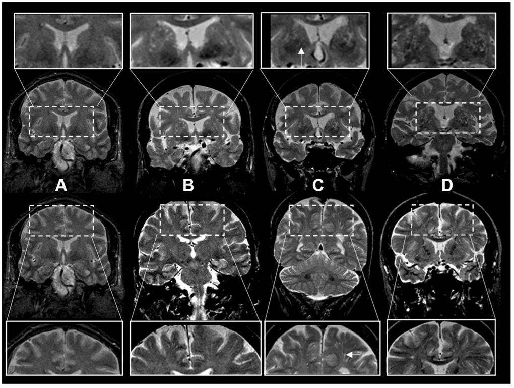
Figure 2.Enlarged perivascular space burden using ratings in coronal T2-weighted MRI sequences. *Top rows: basal ganglia region. Bottom rows: centrum semiovale region. The inserts represent a closer view. White arrows point to individual examples of enlarged perivascular spaces (panel C inserts). †Grades of ePVS burden are based on T2 weighted coronal views. (A) Grade I (0-10), (B) Grade II (10-20) (C) Grade III (20-40) (D) Grade IV (40+).