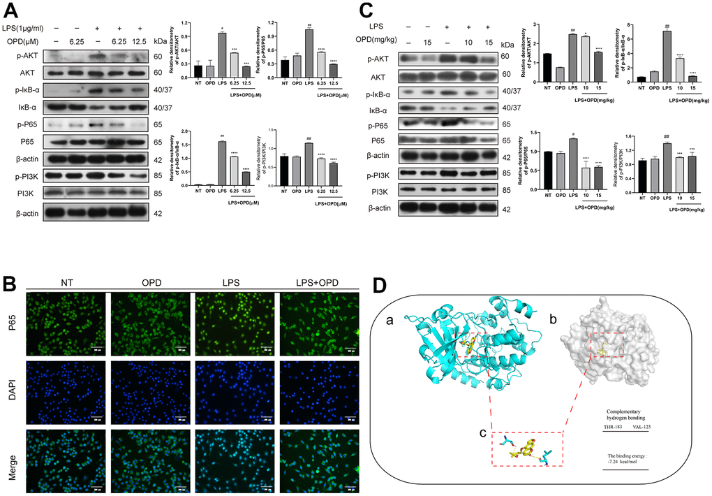
Figure 3.Oxypeucedanin inhibited the activation of the PI3K/AKT/NF-κB signaling pathways in LPS-treated mice and LPS-treated RAW264.7 cells. (A) The cells were pretreated with oxypeucedanin (6.25 and 12.5 μM) for 1 h and then stimulated with LPS for 1 h before the proteins were harvested. Western blotting was used to analyze the protein bands of the PI3K/AKT and NF-κB signaling pathways in RAW264.7 cells (n=5). The quantitative analysis of the protein levels in the AKT and NF-κB signaling pathways is shown. (B) The fluorescence intensity of the translocation of P65 into the nucleus is in RAW264.7 cells. Green indicates P65 proteins, and blue indicates DAPI. Scale bar = 200 μm (n=5). (C) Western blotting was used to analyze the PI3K/AKT and NF-κB signaling pathway proteins in LPS-induced mice. Quantitative analysis of the proteins in the PI3K/AKT and NF-κB signaling pathways (n=3) is shown. (D) The Pdb experiment database was used to download the pdb format of the AKT protein. Pubchem was used to download the sdf structure of oxypeucedanin molecules. AutoDockTools-1.5.6 and PyMOL software were used for molecular docking and data analysis. Yellow represents oxypeucedanin, and the sum can be <0 with statistical significance. (a, b) The surface binding mode of oxypeucedanin and AKT (30w3); (c) OPD binds to AKT (30w3) amino acid residues through hydrogen bonding. The concentrations of OPD in cell and animal experiments were 12.5μM and 15mg/kg, respectively. SEM was used as the error standard for data analysis, and the experiment was repeated three times independently. #p < 0.01 and ##p < 0.0001 compared with No-treatment group; ***p< 0.001 and ****p < 0.0001 compared with the LPS group. LPS: Lipopolysaccharide; OPD: Oxypeucedanin; NT: No-treatment group.