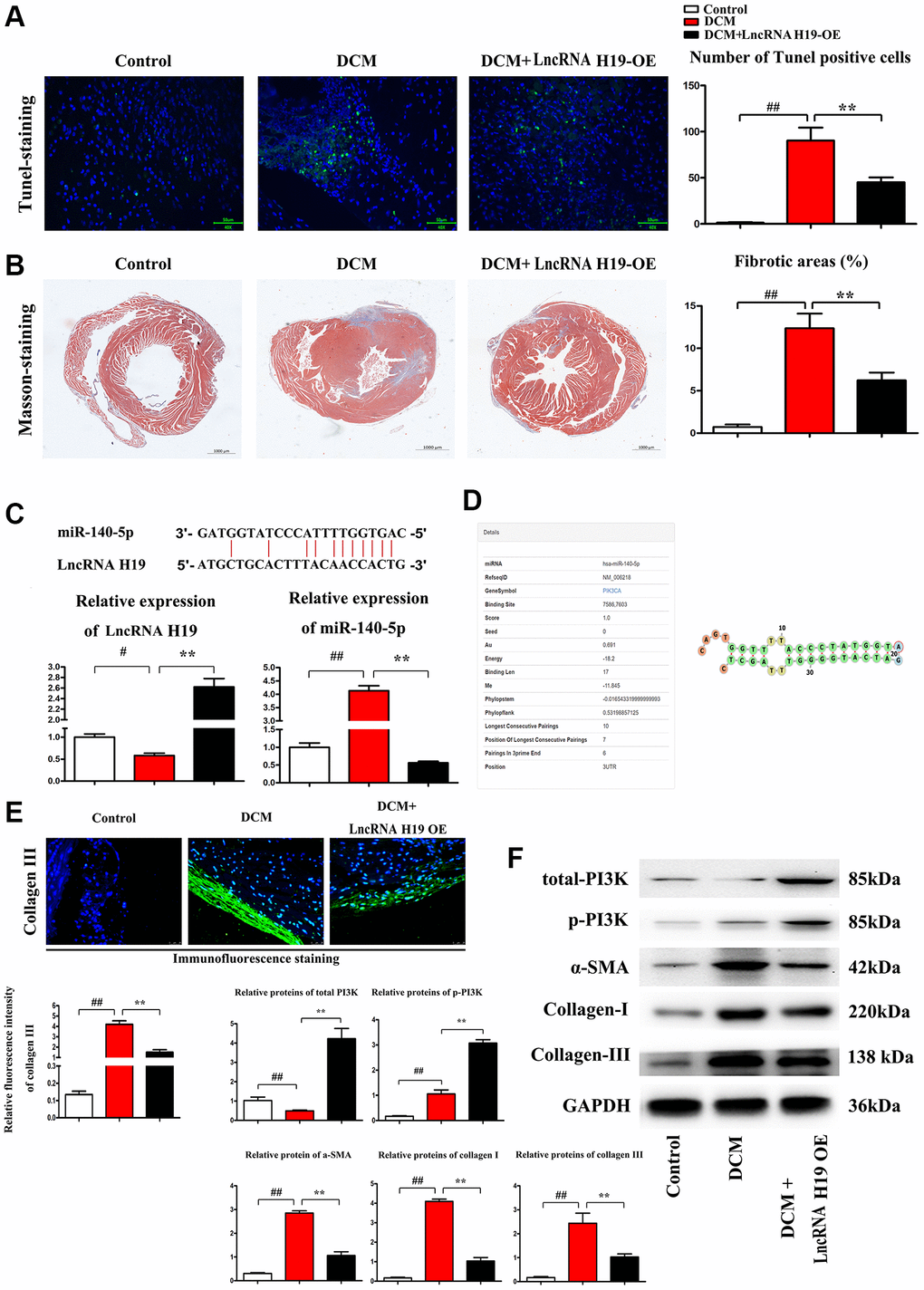
Figure 2.H19 reduced cardiomyocyte apoptosis and cardiac fibrosis in DM. (A) TUNEL staining was used to examine the apoptosis of cardiomyocytes. (B) Masson's trichrome staining was used to investigate the involvement of H19 in cardiac fibrosis in myocardial tissue. (C) The binding site of lncRNA H19 and miR-140-5p and relative expression of lncRNA H19 and miR-140-5p levels were determined by Q-PCR in each group. (D) Binding site of mir-140-5p to PIK3CA. (E) Immunofluorescence intensity of collagen III in each group. (F) The protein levels of p-PI3K, t-PI3K, a-SMA, and collagen-I/III in each group. #P < 0.05; ##P < 0.01 vs. Sham group; *P < 0.05; **P < 0.01 vs. DCM group. between groups.