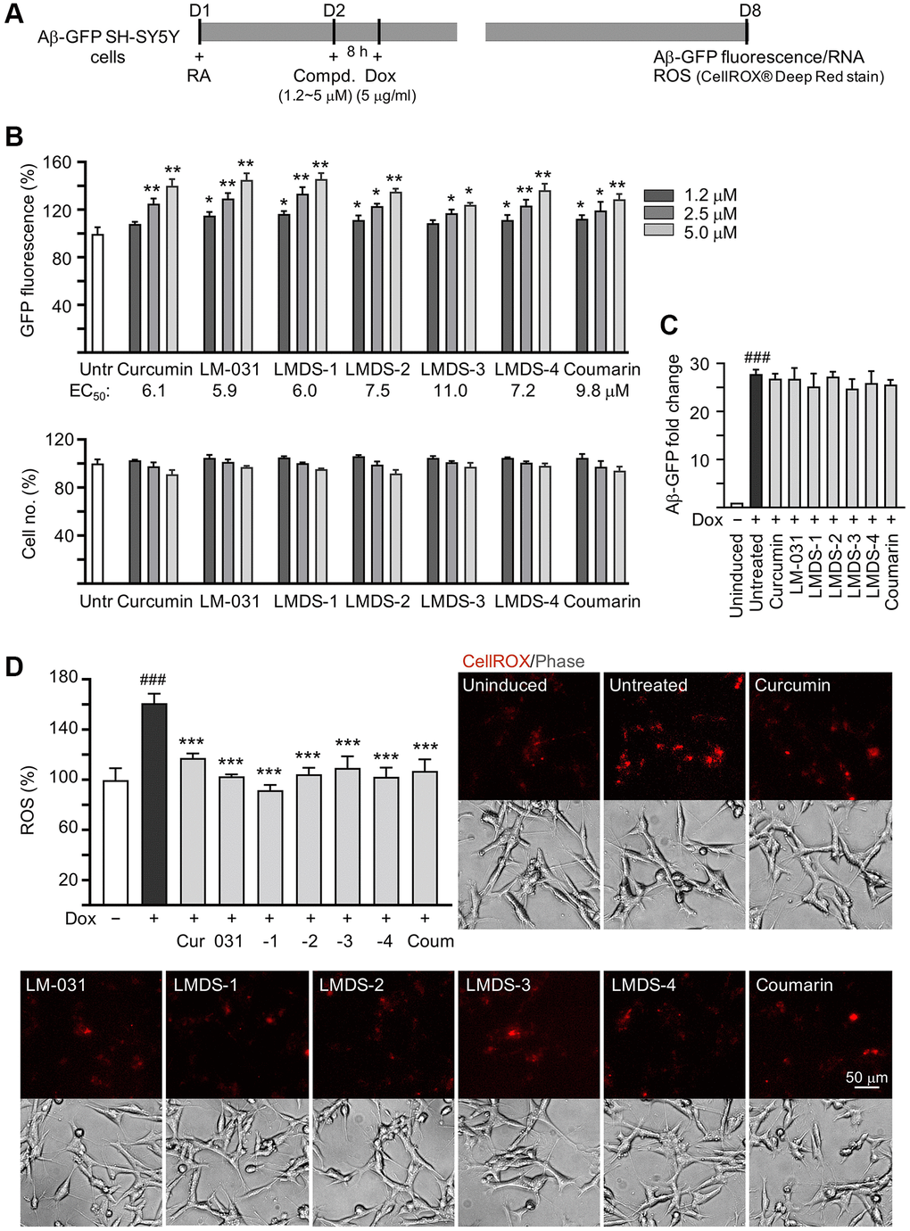
Figure 3.Aβ aggregation and oxidative stress inhibitory effects of LM-031 and analogs in Aβ-GFP SH-SY5Y cells. (A) Experimental flow chart. On day 1, cells were plated with retinoic acid (RA, 10 μM) added to the culture medium. On day 2, curcumin, coumarin, LM-031 or analogs (1.2–5 μM) was added to the cells for 8 h, followed by inducing Aβ-GFP expression with doxycycline (Dox, 5 μg/ml) for 6 days. On day 8, Aβ-GFP fluorescence, Aβ-GFP RNA and ROS (CellROX® Deep Red stain) were measured. (B) Assessment of GFP fluorescence with curcumin, coumarin, LM-031 or analogs (1.2–5 μM) treatment (n = 3), with EC50 values shown below. Shown underneath are cell number analyzed in each treatment. The relative GFP fluorescence/cell number of untreated cells (Untr) was normalized as 100%. (two-tailed Student’s t test; *P < 0.05 and **P < 0.01) (C) Aβ-GFP RNA of Aβ-GFP SH-SY5Y cells untreated or treated with curcumin, coumarin, LM-031 or analogs at 5 μM (n = 3). HPRT1 was used for normalization. (D) Images of CellROX® Deep Red stain (red) and ROS assay of Aβ-GFP cells uninduced, untreated, or treated with curcumin, coumarin, LM-031 or analogs at 5 μM (n = 3). The relative ROS of uninduced cells (Dox-) was normalized (100%). (C, D) P values: comparisons between induced (Dox+) vs. uninduced (Dox-) cells (###P < 0.001), or compound-treated vs. untreated (Dox+) cells (***P < 0.001). (one-way ANOVA with post hoc Tukey test).